Korean J Clin Neurophysiol.
2014 Dec;16(2):89-91. 10.14253/kjcn.2014.16.2.89.
Regrowth of Internal Carotid Artery Aneurysm after Neck Clipping Surgery Presenting with Compressive Optic Neuropathy
- Affiliations
-
- 1Department of Neurology, Kyung Hee University Hospital at Gangdong, Kyung Hee University School of Medicine, Seoul, Korea. azzo73@gmail.com
- KMID: 1737972
- DOI: http://doi.org/10.14253/kjcn.2014.16.2.89
Abstract
- No abstract available.
Figure
-
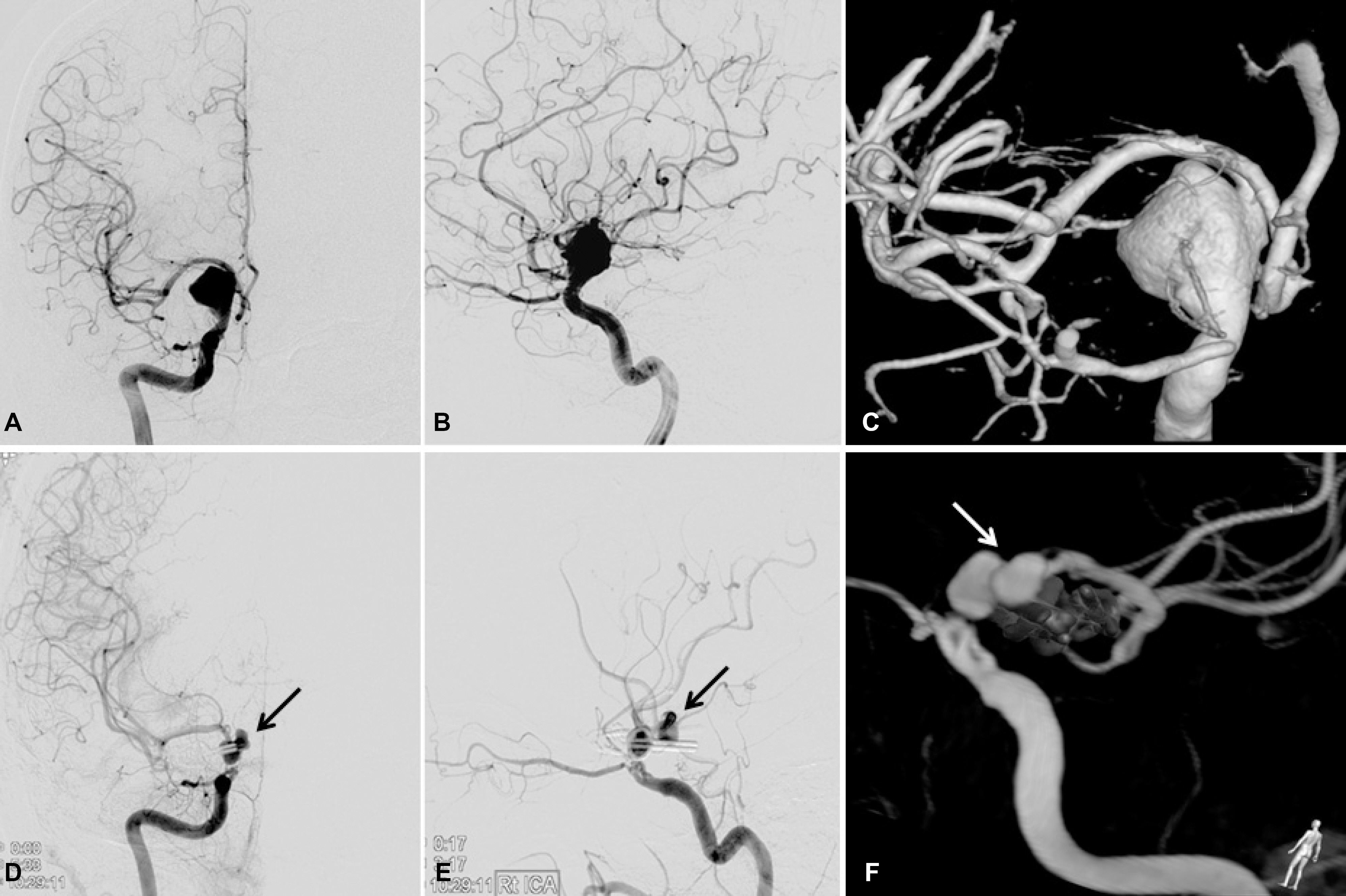
Figure 1. Digital subtraction cerebral angiographic findings. The anterior-posterior (A) and lateral view (B) of the right internal carotid angiogram demonstrates a 15 mm-sized giant aneurysm in the supraclinoid segment of the internal carotid artery (ICA), which bulges out to the lateral direction. Computer-assisted 3D reconstruction of the angiograms (C) provides a clear picture of the dimension of the aneurysm. The anterior-posterior (D) and lateral view (E) of follow up cerebral angiography shows a 7×8×6 mm-sized aneurysm (black arrow) in the supraclinoid segment of the right ICA as well as a clip. The protruding direction of the aneurysm (white arrow) is medial, posterior, and superior to the right ICA (F).
Reference
-
1.Mendez Roberts A., Grimes AL. Enlargement of internal carotid artery aneurysm presenting with severe visual sequela: a case report and anatomy review. Optometry. 2009. 80:76–82.
Article2.Kim SH., Kim SW., Kim BT., Chang JH. A case of intra-cavernous carotid aneurysm presenting with visual loss with no oculomotor disturbance. J Korean Ophthalmol Soc. 2012. 53:486–491.
Article3.Park JY., Koo NK. A giant unruptured aneurysm of distal internal carotid artery presenting with compressive optic neuropathy. J Korean Ophthalmol Soc. 2012. 53:1368–1371.
Article4.el-Beltagy M., Muroi C., Roth P., Fandino J., Imhof HG., Yonekawa Y. Recurrent intracranial aneurysms after successful neck clipping. World Neurosurg. 2010. 74:472–477.5.Peiris JB., Ross Russell RW. Giant aneurysms of the carotid system presenting as visual field defect. J Neurol Neurosurg Psychiatry. 1980. 43:1053–1064.
Article6.David CA., Vishteh AG., Spetzler RF., Lemole M., Lawton MT., Partovi S. Late angiographic follow-up review of surgically treated aneurysms. J Neurosurg. 1999. 91:396–401.
Article7.Tsutsumi K., Ueki K., Morita A., Usui M., Kirino T. Risk of aneurysm recurrence in patients with clipped cerebral aneurysms: results of long-term follow-up angiography. Stroke. 2001. 32:1191–1194.
- Full Text Links
-
- Actions
-
Cited
- CITED
-
- Close
- Share
-
- Similar articles
-
- A Giant Unruptured Aneurysm of Distal Internal Carotid Artery Presenting with Compressive Optic Neuropathy
- Regrowth of Posterior Communicating Artery Aneurysm after 13 Years of the First Clipping: A Case Report
- Monocular Superior Altitudinal Field defect due to Supraclinoid Internal Carotid Artery Aneurysm
- Compressive Optic Neuropathy Caused by Internal Carotid Artery Aneurysm
- Compressive Optic Neuropathy Caused by Internal Carotid Artery Aneurysm Presenting with Concurrent Neuromyelitis Optica
