Patellofemoral Crepitus after Total Knee Arthroplasty: Etiology and Preventive Measures
- Affiliations
-
- 1Colorado Joint Replacement, Denver, CO, USA. kendallslutzky@centura.org
- 2Department of Biomedical Engineering, University of Tennessee, Knoxville, TN, USA.
- 3Department of Bioengineering, University of Denver, Denver, CO, USA.
- 4Department of Orthopaedics, University of Colorado School of Medicine, Denver, CO, USA.
- KMID: 1737626
- DOI: http://doi.org/10.4055/cios.2014.6.1.9
Abstract
- Patellofemoral crepitus and clunk syndrome are infrequent, yet troublesome complications of total knee arthroplasty with a reported incidence of 0%-18%. They are primarily associated with implantation of posterior cruciate substituting designs. These entities are the result of peripatellar fibrosynovial hyperplasia at the junction of the superior pole of the patella and the distal quadriceps tendon which becomes entrapped within the superior aspect of the intercondylar box of the femoral component during knee flexion. When the knee extends, a crepitant sensation occurs as the fibrosynovial tissue exits the intercondylar box. Numerous etiologies have been proposed such as femoral component designs with a high intercondylar box ratio, previous knee surgery, reduced patellar tendon length, thinner patellar components, reduced patella-patellar component composite thickness, and smaller femoral components. Preventative measures include choice of femoral components with a reduced intercondylar box ratio, use of thicker patellar components, avoidance of over-resection of the patella, and debridement of the fibrosynovial tissue at the time of knee arthroplasty. Most patients with crepitus are unaware of the problem or have minimal symptoms so that no treatment is required. If significant disability is incurred, symptoms can be eliminated in a high percentage of patients with arthroscopic debridement of the fibrosynovial hyperplasia.
MeSH Terms
Figure
-
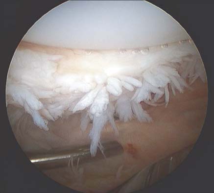
Fig. 1 Arthroscopic view of fibro-synovial hyperplasia at the junction of the superior pole of the patella and the distal quadriceps tendon in a patient with symptomatic patellar crepitus.
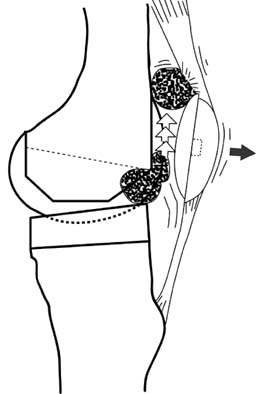
Fig. 2 Schematic drawing of the pathophysiology of patellar clunk syndrome. A discrete fibrosynovial nodule becomes entrapped within the intercondylar box of the posterior stabilized femoral component during flexion and is subsequently released when the knee is extended to within 30-45 degrees of full extension.

Fig. 3 Photograph of a cruciate retaining (left) and posterior stabilized (right) femoral components, demonstrating a higher transition height from the trochlear groove to the intercondylar box in the posterior stabilized femoral component.
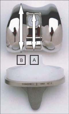
Fig. 4 Photograph of a posterior stabilized femoral component demonstrating the intercondylar box ratio, defined as the height of the intercondylar box divided by the anterior-posterior height of the femoral component.

Fig. 5 Three posterior stabilized (PS) femoral components with different intercondylar box geometries. The design with the greatest intercondylar box height and narrowest intercondylar box width demonstrated the highest incidence of synovial entrapment (AMK Congruency; Depuy, Warsaw, IN, USA). Reprint from Pollock et al.17) with permission from The Journal of Bone and Joint Surgery, Inc.
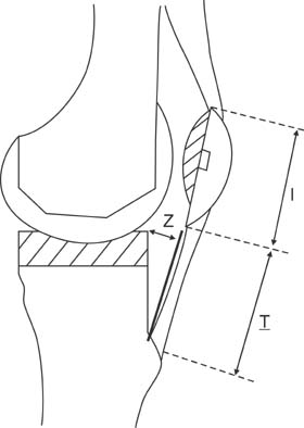
Fig. 6 Diagram demonstrating the Insall-Salvati ratio (T/I) and the perpendicular distance from the upper anterior corner of the tibial tray to the patellar tendon ([Z]: measuring the anterior posterior relationship of the tibial tray relative to the extensor mechanism). Reprint from Yau et al.16) with permission from Elsevier.
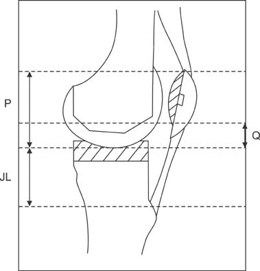
Fig. 7 Diagram demonstrating the joint line (JL), patellar button height (Q), and position of the proximal pole of the patella with reference to distal end of the femoral prosthesis (P). Reprint from Yau et al.16) with permission from Elsevier.

Fig. 8 Composite contract area within 2 mm of intercondylar notch (up to 120°) with original ligament and changes due to variation in patellar tendon length (alta and baja), flexion-extension alignment of the femoral component, join line and patellar button size. Reprint from Hoops et al.31) with permissison from John Wiley & Sons, Inc.
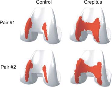
Fig. 9 Cumulative tendo-femoral contact patches in matched-control pairs of patients with and without patellofemoral crepitus.

Fig. 10 Cumulative tendo-femoral contact patches in a patient with patellofemoral crepitus with potential changes in femoral flexion and joint line.
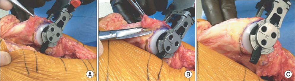
Fig. 11 Intraoperative photographs of the posterior aspect of the distal quadriceps tendon, demonstating synovial proliferation at the border of the superior pole of the patella and distal quadriceps tendon (A), and its removal (B, C).
Cited by 1 articles
-
Noise around the Knee
Sang Jun Song, Cheol Hee Park, Hu Liang, Sang Jun Kim
Clin Orthop Surg. 2018;10(1):1-8. doi: 10.4055/cios.2018.10.1.1.
Reference
-
1. Wing CK, Kwok-Hing C. Sixteen years' result of posterior-stabilized TKA. J Knee Surg. 2012; 25(3):245–248.2. Thadani PJ, Vince KG, Ortaaslan SG, Blackburn DC, Cudiamat CV. Ten- to 12-year followup of the Insall-Burstein I total knee prosthesis. Clin Orthop Relat Res. 2000; (380):17–29.3. Meftah M, Ranawat AS, Ranawat CS. The natural history of anterior knee pain in 2 posterior-stabilized, modular total knee arthroplasty designs. J Arthroplasty. 2011; 26(8):1145–1148.4. Figgie HE 3rd, Goldberg VM, Heiple KG, Moller HS 3rd, Gordon NH. The influence of tibial-patellofemoral location on function of the knee in patients with the posterior stabilized condylar knee prosthesis. J Bone Joint Surg Am. 1986; 68(7):1035–1040.5. Insall JN, Lachiewicz PF, Burstein AH. The posterior stabilized condylar prosthesis: a modification of the total condylar design: two to four-year clinical experience. J Bone Joint Surg Am. 1982; 64(9):1317–1323.6. Hozack WJ, Rothman RH, Booth RE Jr, Balderston RA. The patellar clunk syndrome: a complication of posterior stabilized total knee arthroplasty. Clin Orthop Relat Res. 1989; (241):203–208.7. Niikura T, Tsumura N, Tsujimoto K, Yoshiya S, Kurosaka M, Shiba R. Patellar clunk syndrome after TKA with cruciate retaining design: a report of two cases. Orthopedics. 2008; 31(1):90.8. Sringari T, Maheswaran SS. Patellar clunk syndrome in patellofemoral arthroplasty: a case report. Knee. 2005; 12(6):456–457.9. Beight JL, Yao B, Hozack WJ, Hearn SL, Booth RE Jr. The patellar "clunk" syndrome after posterior stabilized total knee arthroplasty. Clin Orthop Relat Res. 1994; (299):139–142.10. Clarke HD, Fuchs R, Scuderi GR, Mills EL, Scott WN, Insall JN. The influence of femoral component design in the elimination of patellar clunk in posterior-stabilized total knee arthroplasty. J Arthroplasty. 2006; 21(2):167–171.11. Fukunaga K, Kobayashi A, Minoda Y, Iwaki H, Hashimoto Y, Takaoka K. The incidence of the patellar clunk syndrome in a recently designed mobile-bearing posteriorly stabilised total knee replacement. J Bone Joint Surg Br. 2009; 91(4):463–468.12. Ip D, Ko PS, Lee OB, Wu WC, Lam JJ. Natural history and pathogenesis of the patella clunk syndrome. Arch Orthop Trauma Surg. 2004; 124(9):597–602.13. Lonner JH, Jasko JG, Bezwada HP, Nazarian DG, Booth RE Jr. Incidence of patellar clunk with a modern posterior-stabilized knee design. Am J Orthop (Belle Mead NJ). 2007; 36(10):550–553.14. Ranawat AS, Ranawat CS, Slamin JE, Dennis DA. Patellar crepitation in the P.F.C. sigma total knee system. Orthopedics. 2006; 29:9 Suppl. S68–S70.15. Schroer WC, Diesfeld PJ, Reedy ME, LeMarr A. Association of increased knee flexion and patella clunk syndrome after mini-subvastus total knee arthroplasty. J Arthroplasty. 2009; 24(2):281–287.16. Yau WP, Wong JW, Chiu KY, Ng TP, Tang WM. Patellar clunk syndrome after posterior stabilized total knee arthroplasty. J Arthroplasty. 2003; 18(8):1023–1028.17. Pollock DC, Ammeen DJ, Engh GA. Synovial entrapment: a complication of posterior stabilized total knee arthroplasty. J Bone Joint Surg Am. 2002; 84(12):2174–2178.18. Ip D, Wu WC, Tsang WL. Comparison of two total knee prostheses on the incidence of patella clunk syndrome. Int Orthop. 2002; 26(1):48–51.19. Maloney WJ, Schmidt R, Sculco TP. Femoral component design and patellar clunk syndrome. Clin Orthop Relat Res. 2003; (410):199–202.20. Anderson MJ, Becker DL, Kieckbusch T. Patellofemoral complications after posterior-stabilized total knee arthroplasty: a comparison of 2 different implant designs. J Arthroplasty. 2002; 17(4):422–426.21. Shoji H, Shimozaki E. Patellar clunk syndrome in total knee arthroplasty without patellar resurfacing. J Arthroplasty. 1996; 11(2):198–201.22. Frye BM, Floyd MW, Pham DC, Feldman JJ, Hamlin BR. Effect of femoral component design on patellofemoral crepitance and patella clunk syndrome after posterior-stabilized total knee arthroplasty. J Arthroplasty. 2012; 27(6):1166–1170.23. Dennis DA, Kim RH, Johnson DR, Springer BD, Fehring TK, Sharma A. The John Insall Award: control-matched evaluation of painful patellar Crepitus after total knee arthroplasty. Clin Orthop Relat Res. 2011; 469(1):10–17.24. Dajani KA, Stuart MJ, Dahm DL, Levy BA. Arthroscopic treatment of patellar clunk and synovial hyperplasia after total knee arthroplasty. J Arthroplasty. 2010; 25(1):97–103.25. Koh YG, Kim SJ, Chun YM, Kim YC, Park YS. Arthroscopic treatment of patellofemoral soft tissue impingement after posterior stabilized total knee arthroplasty. Knee. 2008; 15(1):36–39.26. Wong JW, Yau PW, Chiu PK. Arthroscopic treatment of patellar symptoms in posterior stabilized total knee replacement. Int Orthop. 2002; 26(4):250–252.27. Takahashi M, Miyamoto S, Nagano A. Arthroscopic treatment of soft-tissue impingement under the patella after total knee arthroplasty. Arthroscopy. 2002; 18(4):E20.28. Lucas TS, DeLuca PF, Nazarian DG, Bartolozzi AR, Booth RE Jr. Arthroscopic treatment of patellar clunk. Clin Orthop Relat Res. 1999; (367):226–229.29. Messieh M. Management of patellar clunk under local anesthesia. J Arthroplasty. 1996; 11(2):202–203.30. Vernace JV, Rothman RH, Booth RE Jr, Balderston RA. Arthroscopic management of the patellar clunk syndrome following posterior stabilized total knee arthroplasty. J Arthroplasty. 1989; 4(2):179–182.31. Hoops HE, Johnson DR, Kim RH, et al. Control-matched computational evaluation of tendo-femoral contact in patients with posterior-stabilized total knee arthroplasty. J Orthop Res. 2012; 30(9):1355–1361.
- Full Text Links
-
- Actions
-
Cited
- CITED
-
- Close
- Share
-
- Similar articles
-
- Total Knee Arthroplasty without Patellar Resurfacing in Moderate to Severe Patellofemoral Arthritis
- The Effect of Patellofemoral Overstuffing in Total Knee Arthroplasty
- Radiologic Patellar Change and Clinical Results of Total Knee Arthroplasty without Patella Resurfacing
- Noise around the Knee
- Functional and Radiological Outcomes of Patella Retaining Total Knee Arthroplasty: Minimun 5-Year Follow-up Results
