MR Imaging in a Child with Scurvy: a Case Report
- Affiliations
-
- 1Department of Radiology, College of Medicine, Inha University, Incheon, Korea. swpark88@inha.ac.kr
- 2Department of Pediatrics, College of Medicine, Inha University, Incheon, Korea.
- 3Department of Orthopeadics, College of Medicine, Inha University, Incheon, Korea.
- KMID: 1734294
- DOI: http://doi.org/10.3348/kjr.2007.8.5.443
Abstract
- Scurvy is very rare disease in industrialized societies. Nevertheless, it still exists in higher risk groups including economically disadvantaged populations with poor nutrition, such as the elderly and chronic alcoholics. The incidence of scurvy in the pediatric population is very low. This study reports a case of scurvy in a 5-year-old girl with cerebral palsy and developmental delay based on MRI findings.
Keyword
MeSH Terms
-
Ascorbic Acid/blood/therapeutic use
Bone Diseases, Metabolic/etiology
Cerebral Palsy/complications
Child, Preschool
Cholecalciferol/blood
Developmental Disabilities/complications
Drainage
Female
Femur/pathology/radionuclide imaging/surgery
Fever/etiology
Follow-Up Studies
Hematoma/diagnosis/etiology/surgery
Humans
Knee/radiography
Magnetic Resonance Imaging/*methods
Muscle Weakness/etiology
Rare Diseases
Scurvy/complications/*diagnosis/drug therapy
Thigh/pathology
Vitamins/therapeutic use
Figure
-
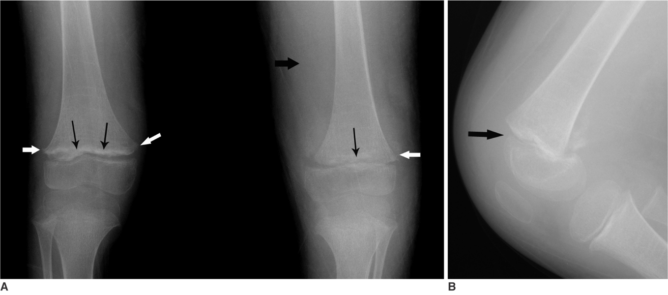
Fig. 1 A. Anteroposterior radiograph of both knees shows a thick sclerotic metaphyseal line (thin black arrows) above a widened physis and small beaklike excrescences (white arrows) at the metaphysis in both femora. Soft tissue bulging is noted (thick black arrow). B.Lateral radiograph of the left knee shows a disruption of the alignment of the distal femoral physis (arrow).

Fig. 2 MRI of the left thigh performed at the first hospital day. A. Coronal T2-weighted image shows a diffuse bone marrow signal change (black arrows) of the femur shaft with a large amount of subperiosteal fluid collection (arrowhead) and displacement of the distal epiphysis (white arrow). B.Axial T2-weighted image shows a fluid-fluid level in the subperiosteal fluid collection (black arrow). The surrounding vastus and hamstring muscles also show high intensity signal lesions (white arrows).
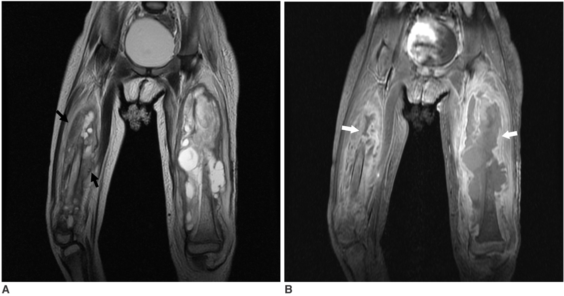
Fig. 3 Follow up MR images of both thighs. A. A coronal T2-weighted image shows a larger subperiosteal hematoma at the left femur and a new subperiosteal hematoma (arrows) with signal change of bone marrow of the right femur. B.A coronal contrast enhanced fat suppression T1-weighted image shows moderate enhancement of the periosteum and adjacent soft tissue (white arrows).
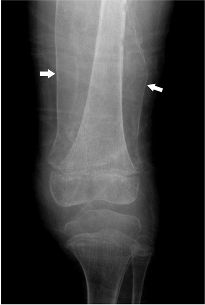
Fig. 4 The radiograph performed six weeks after vitamin C supplementation. Large shells of periosteal bone (arrows) are present at the left femur. There was improvement of the metaphyseal sclerotic line and widened physis compared to the initial radiograph.
Reference
-
1. Olmedo JM, Yiannias JA, Windgassen EB, Gornet MK. Scurvy: a disease almost forgotten. Int J Dermatol. 2006. 45:909–913.2. Fain O. Musculoskeletal manifestations of scurvy. Joint Bone Spine. 2005. 72:124–128.3. Weinstein M, Babyn P, Zlotkin S. An orange a day keeps the doctor away: scurvy in the year 2000. Pediatrics. 2001. 108:E55.4. Halligan TJ, Russel NG, Dunn WJ, Caldroney SJ, Skelton TB. Identification and treatment of scurvy: a case report. Oral Surg, Oral Med, Oral Path, Oral Radiol, and Endod. 2005. 100:688–692.5. Pimentel L. Scurvy: historical review and current diagnostic approach. Am J Emerg Med. 2003. 21:328–332.6. Resnick D. Hypervitaminosis and hypovitaminosis. Diganosis of bone and joint disorders. 2002. 4th ed. Philadelphia: WB Saunders Company;3456–3464.
- Full Text Links
-
- Actions
-
Cited
- CITED
-
- Close
- Share
-
- Similar articles
-
- A Case of Scurvy Mimics Cutaneous Vasculitis
- Undifferentiated Embryonal Sarcoma of Liver in Child
- Arterial Spin Labelling Perfusion, Proton MR Spectroscopy and Susceptibility-Weighted MR Findings of Acute Necrotizing Encephalopathy: a Case Report
- Probable Cases of Scurvy in Subadults Crania from Archaeological Sites, South Korea
- A case of Scurvy
