Mazabraud's Syndrome Coexisting with a Uterine Tumor Resembling an Ovarian Sex Cord Tumor (UTROSCT): a Case Report
- Affiliations
-
- 1Department of Radiology, Eskisehir Osmangazi University, Turkey. cuneytcalisir_72@yahoo.com
- 2Department of Orthopaedics and Traumatology, Eskisehir Osmangazi University, Turkey.
- 3Department of Pathology, Eskisehir Osmangazi University, Turkey.
- KMID: 1734293
- DOI: http://doi.org/10.3348/kjr.2007.8.5.438
Abstract
- The association of intramuscular myxoma and fibrous dysplasia is a rare disease known as Mazabraud's syndrome. We present a case of Mazabraud's syndrome coexisting with a uterine tumor and resembling an ovarian sex cord tumor (UTROSCT). This uterine tumor showed a high mitotic index and cytological atypia. To the best of our knowledge, the coexistence of the two different entities has not been reported in the literature.
Keyword
MeSH Terms
-
Aged
Biopsy
Buttocks/pathology/surgery/ultrasonography
Diagnosis, Differential
Female
Fibrous Dysplasia, Monostotic/complications/*diagnosis/surgery
Humans
Magnetic Resonance Imaging
Myxoma/complications/*diagnosis/surgery
Ovarian Neoplasms/*diagnosis
Rare Diseases
Sex Cord-Gonadal Stromal Tumors/*diagnosis
Syndrome
Uterine Neoplasms/complications/*diagnosis/surgery
Figure
-
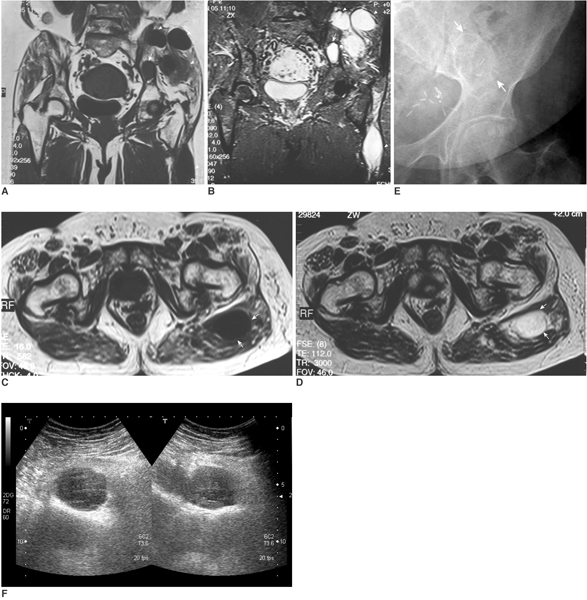
Fig. 1 A, B. Coronal T1-weighted (A) and coronal STIR MR images (B) demonstrate an area of signal abnormality within the left ilium. The region appears with low signal intensity on T1 and high signal intensity on STIR relative to adult yellow marrow, consistent with fibrous dysplasia (arrow). Also identified are multiple intramuscular masses within the left gluteal musculature and left thigh, which appear with low signal intensity on T1-weighted images and high signal intensity on STIR images relative to muscle, consistent with myxomas (arrowheads). C, D. An axial T1-weighted image (C) Axial T2-weighted MR images (D) demonstrate an oval, sharply defined mass in the left gluteus maximus, which has a low signal intensity on T1 and high signal intensity on T2 with homogeneity in both signals (arrows). E. An anteroposterior radiograph of the left hip demonstrates a well-defined oval osteolytic lesion with a thin, sclerotic rim within the left ilium (arrows). F. Ultrasonography of the left gluteal musculature shows a heterogeneous, solid, hypoechoic, lobulated intramuscular tumor with multiple small-sized fluid filled cystic areas.
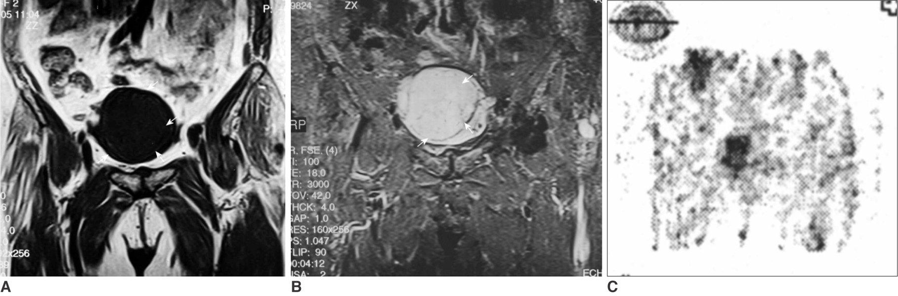
Fig. 2 A. Coronal T1 weighted MR image shows a solid mass within the enlarged endometrial cavity. The tumor has regular margins and homogenous low signal intensity (arrows). B. A coronal STIR MR image of the pelvis. The uterine mass is observed as a homogenous area of high signal intensity in the uterus (arrows). There is no evidence of necrotic areas within the lesion. C. FDG-PET image. Accumulated FDG is seen in the uterine lesion.
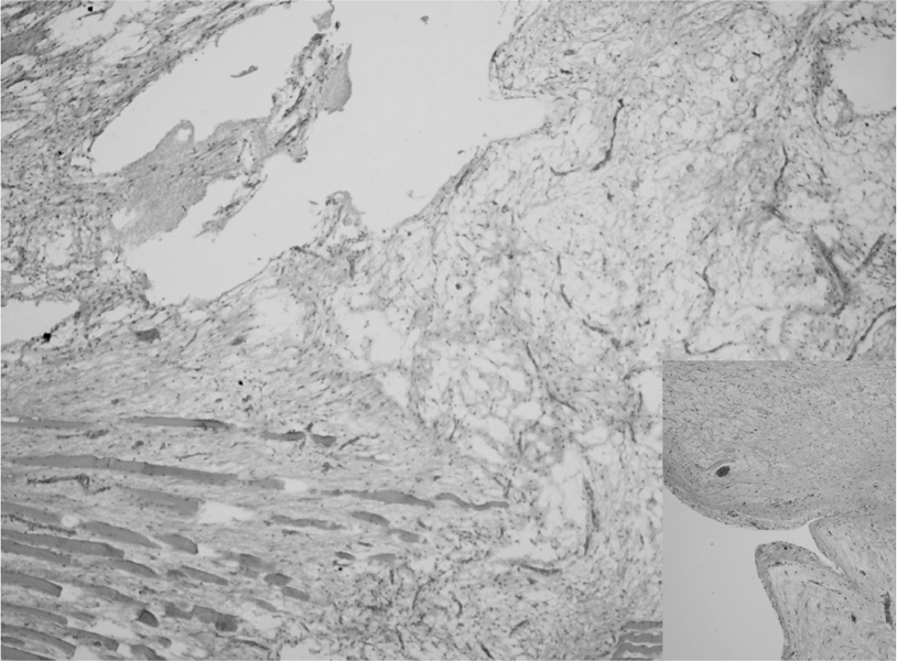
Fig. 3 Intramuscular myxoma. A hypocellular tumor extending into the striated muscle is composed of spindle and stellate cells. The stroma is myxoid in appearance and showed cystic degeneration (Hematoxylin & Eosin staining, × 4). Insert: A cystic space lined with fibrous tissue and tumor cells in its wall (Hematoxylin & Eosin staining, × 10).
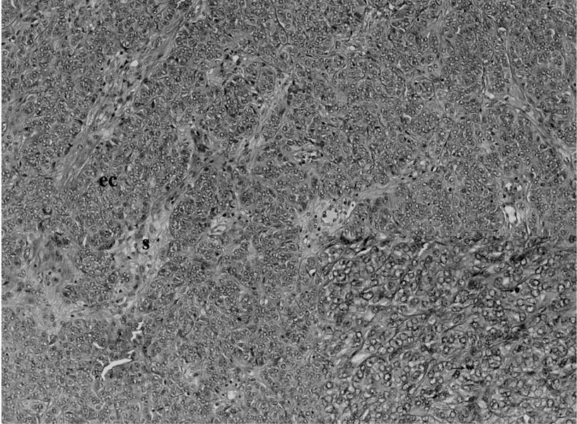
Fig. 4 The uterine tumor consisted of the cell groups in epithelioid appearance (ec). They were arranged in cords and trabecula resembling sex cord structures in a scanty fibrous stroma (s) (Hematoxylin & Eosin staining, × 10). Insert: Higher magnification of the tumor cells shows cytological atypia and mitosis (arrows) (Hematoxylin & Eosin staining, × 40).
Reference
-
1. Court-Payen M, Ingemann JL, Bjerregaard B, Schwarz LG, Skjoldbye B. Intramuscular myxoma and fibrous dysplasia of bone-Mazabraud's syndrome. Acta Radiol. 1997. 38:368–371.2. Clement PB, Scully RE. Uterine tumors resembling ovarian sex-cord tumors. Am J Clin Pathol. 1976. 66:512–525.3. Hendrickson MR, Longacre TA, Kempson RL. Sternberg SS, editor. The Uterine Corpus. Diagnostic surgical pathology. 1999. 3rd ed. Philadelphia: Williams & Wilkins;2269–2270.4. Iwasko N, Steinbach LS, Disler D, Pathria M, Hottya GA, Kattapuram S, et al. Imaging findings in Mazabraud's syndrome: seven new cases. Skeletal Radiol. 2002. 31:81–87.5. Mazabraud A, Girard J. A peculiar case of fibrous dysplasia with osseous and tendinous localizations. Rev Rhum Mal Osteoartic. 1957. 24:652–659.6. Wirth WA, Leavitt D, Enzinger FM. Multiple intramuscular myxomas. Another extraskeletal manifestation of fibrous dysplasia. Cancer. 1971. 27:1167–1173.7. Totty WG, Murphy WA, Lee JK. Soft tissue tumors: MR imaging. Radiology. 1986. 160:135–141.8. Prayson MA, Leeson MC. Soft-tissue myxomas and fibrous dysplasia of bone. A case report and review of the literature. Clin Orthop Relat Res. 1993. 291:222–228.9. Suzuki C, Matsumoto T, Fukunaga M, Itoga T, Furugen Y, Kurosaki Y, et al. Uterine tumors resembling sex-cord tumors producing parathyroid hormone-related protein of the uterine cervix. Pathol Int. 2002. 52:164–168.10. Okada S, Uchiyama F, Ohaki Y, Kamoi S, Kawamura T, Kumazaki T. MRI findings of a case of uterine tumor resembling ovarian sex-cord tumors coexisting with endometrial adenoacanthoma. Radiat Med. 2001. 19:151–153.
- Full Text Links
-
- Actions
-
Cited
- CITED
-
- Close
- Share
-
- Similar articles
-
- A case of uterine tumor resembling ovarian sex-cord tumor with clinical review
- A case of uterine tumor resembling ovarian sex-cord tumor
- Two Cases of Uterine Tumors Resembling Ovarian Sex-cord Tumors: Rare Case of Uterine Tumor
- Successful delivery after conservative resectoscopic surgery in a patient with a uterine tumor resembling ovarian sex cord tumor with myometrial invasion
- Uterine Tumor Resembling Ovarian Sex-Cord Tumor: A case report
