Intracranial Dural Metastasis of Ewing's Sarcoma: a Case Report
- Affiliations
-
- 1Department of Radiology, the Research Institute of Radiological Science, Yonsei University College of Medicine, Seoul, Korea. eungykim@yuhs.ac
- 2Department of Neurosurgery, Yonsei University College of Medicine, Seoul, Korea.
- 3Department of Pathology, Yongdong Severance Hospital, Yonsei University College of Medicine, Seoul, Korea.
- KMID: 1734278
- DOI: http://doi.org/10.3348/kjr.2008.9.1.76
Abstract
- Although intracranial dural metastasis of Ewing's sarcoma is a very rare finding, its imaging characteristics are similar to those of its primary form in the central nervous system. Thus, this tumor must be considered in the differential diagnosis of extra-axial dural masses.
Keyword
MeSH Terms
Figure
-
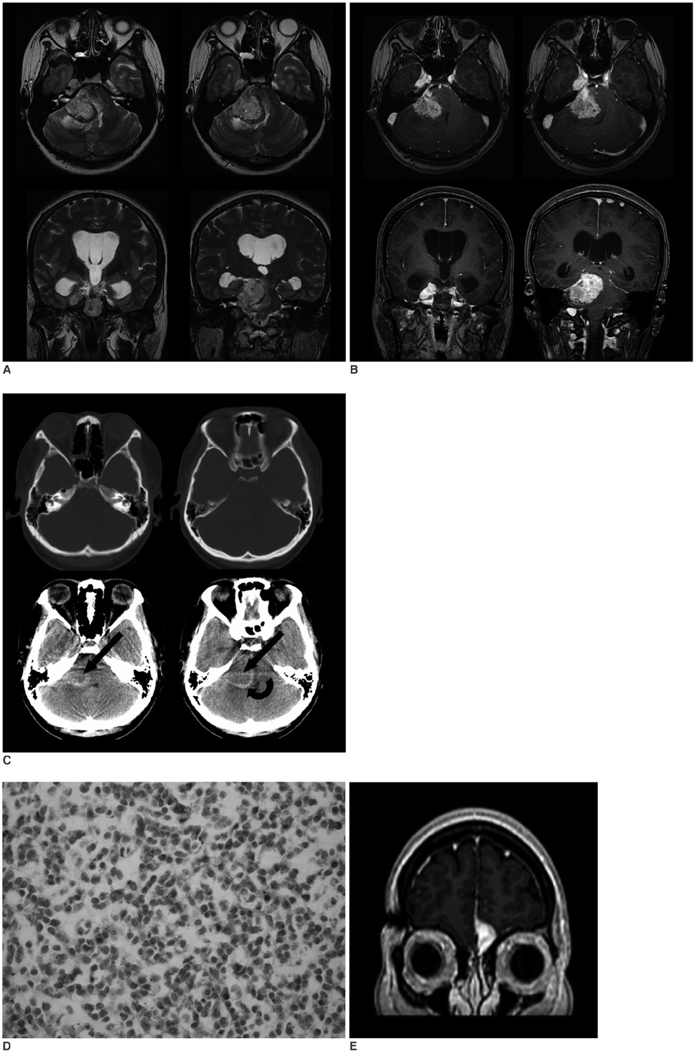
Fig. 1 The T2-weighted (A) and postcontrast T1-weighted images (B) show a mass in the right cerebellopontine angle cistern with extension into the ipsilateral Meckel's cave. The mass shows a slightly heterogeneous lobulated contour with similar or slightly higher signal intensity than the cortex on the T2-weighted images, with heterogeneous enhancement. Edema is noted in the pons and cerebellum adjacent to the mass. Noncontrast CT shows spots of subtle high-attenuation within the mass (arrows), suggesting the possibility of calcification or hemorrhage. Also seen is a high-attenuation curvilinear area between the mass and the cerebellum (curved arrow), which was determined to be fibrosis on the pathologic examination (C). The ipsilateral internal auditory canal and petrous bone are free of tumor on both the CT and MRI (A-C). Immunohistochemistry demonstrates the MIC-2 antigen expression (D). Follow-up MRI reveals a new enhancing lesion abutting the anterior falx cerebri and left fovea ethmoidalis, and this suggests metastasis (E).
Reference
-
1. Pizo P, Poplack D. Ewing's sarcoma of bone and soft tissue: principles and practice of pediatric oncology. 1996. 3rd ed. Philadelphia: JB Lippincott;840–841.2. Postovsky S, Ash S, Ramu IN, Yaniv Y, Zaizov R, Futerman B, et al. Central nervous system involvement in children with sarcoma. Oncology. 2003. 65:118–124.3. Shuper A, Cohen IJ, Mor C, Ash S, Kornreich L, Zaizov R. Metastatic brain involvement in Ewing family of tumors in children. Neurology. 1998. 51:1336–1338.4. Li WY, Brock P, Saunders DE. Imaging characteristics of primary cranial Ewing sarcoma. Pediatr Radiol. 2005. 35:612–618.5. Mehta Y, Hendrickson FR. CNS involvement in Ewing's sarcoma. Cancer. 1974. 33:859–862.6. Marciani MG, Stefani N, Peroni L, Stefanini F, Tarantino U, Gigli GL, et al. Intracerebral metastasis in Ewing's sarcoma. Acta Neurol Belg. 1990. 90:149–156.7. Meyer PC, Reah TG. Secondary neoplasms of the central nervous system and meninges. Br J Cancer. 1953. 7:438–448.8. Kleinschmidt-DeMasters BK. Dural metastases. A retrospective surgical and autopsy series. Arch Pathol Lab Med. 2001. 125:880–887.9. Pekala JS, Gururangan S, Provenzale JM, Mukundan S Jr. Central nervous system extraosseous Ewing sarcoma: radiologic manifestations of this newly defined pathologic entity. AJNR Am J Neuroradiol. 2006. 27:580–583.10. Dirks PB, Harris L, Hoffman HJ, Humphreys RP, Drake JM, Rutka JT. Supratentorial primitive neuroectodermal tumors in children. J Neurooncol. 1996. 29:75–84.
