Korean J Radiol.
2008 Feb;9(1):54-58. 10.3348/kjr.2008.9.1.54.
In Utero Development of the Fetal Gall Bladder in the Korean Population
- Affiliations
-
- 1Department of Radiology, Cheil General Hospital & Women's Healthcare Center, Kwandong University College of Medicine, Seoul, Korea.
- 2Department of Radiology, Seoul National University College of Medicine, and the Institute of Radiation Medicine, Seoul, Korea. radjycho@radiol.snu.ac.kr
- KMID: 1734275
- DOI: http://doi.org/10.3348/kjr.2008.9.1.54
Abstract
OBJECTIVE
To provide reference ranges of the fetal gall bladder in the Korean population. MATERIALS AND METHODS: Fetal gall bladder development was evaluated in well-dated, non-anomalous fetuses in the Korean population between February and April 2003 and the visualization rate and reference values were determined from the obtained data. RESULTS: The visualization rate of the fetal gall bladder increased as gestation advanced to a plateau above 90%, which was maintained between 16 and 34 weeks. The measured parameters from the fetal gall bladder had a significant positive relationship with gestational age (p = 0.000 for all cases), and the correlation of length and area with the gestational age (r = 0.741 and r = 0.690, respectively) was better than the correlation of width, height, and volume with gestational age. The repeatability coefficients and coefficients of variation between the two operators were 5.56 mm and 12.9% for the length and 344.11 mm(2) and 33.52% for the area. The median length of the fetal gall bladder in the Korean population was not significantly different from the mean length of gall bladders in the Caucasian and African-American populations (p = 0.915). CONCLUSION: We have provided reference values for the fetal gall bladder throughout the gestation period in the Korean population.
Keyword
MeSH Terms
Figure
-
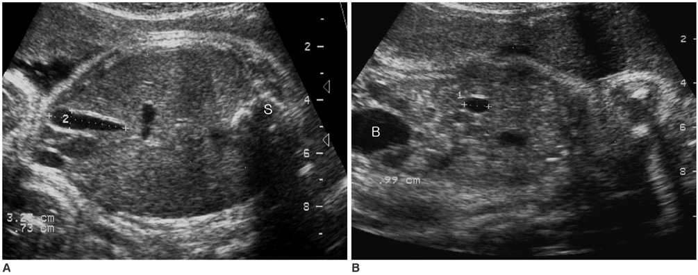
Fig. 1 Sonogram demonstrating fetal gall bladder length and width (A) and height (B). The height was obtained by rotating the transducer 90 degree from plane (A) showing maximal length and width of the gall bladder. The caliper placement for measurement of the gall bladder is demonstrated. S = spine, B = bladder

Fig. 2 Visualization rate of the fetal gall bladder according to gestational age.
Reference
-
1. Chan L, Rao BK, Jiang Y, Endicott B, Wapner RJ, Reece EA. Fetal gallbladder growth and development during gestation. J Ultrasound Med. 1995. 14:421–425.2. Goldstein I, Tamir A, Weisman A, Jakobi P, Copel JA. Growth of the fetal gall bladder in normal pregnancies. Ultrasound Obstet Gynecol. 1994. 4:289–293.3. Jones KL. Jones KL, editor. Chromosomal abnormality syndrome. Smith's recognizable patterns of human malformations. 1988. Philadelphia: Saunders;10–25.4. Bland JM, Altman DG. Measuring agreement in method comparison studies. Stat Methods Med Res. 1999. 8:135–160.5. Bromley B, Frigoletto FD Jr, Harlow BL, Evans JK, Benacerraf BR. Biometric measurements in fetuses of different race and gender. Ultrasound Obstet Gynecol. 1993. 3:395–402.6. Davis RO, Cutter GR, Goldenberg RL, Hoffman HJ, Cliver SP, Brumfield CG. Fetal biparietal diameter, head circumference, abdominal circumference and femur length. A comparison by race and sex. J Reprod Med. 1993. 38:201–206.7. Jacquemyn Y, Sys SU, Verdonk P. Fetal transverse cerebellar diameter in different ethnic groups. J Perinat Med. 2000. 28:14–19.8. Moon MH, Cho JY, Lee YM, Lee YH, Yang JH, Kim MY, et al. Nasal bone length at 11-14 weeks of pregnancy in the Korean population. Prenat Diagn. 2006. 26:524–527.9. Bronshtein M, Weiner Z, Abramovici H, Filmar S, Erlik Y, Blumenfeld Z. Prenatal diagnosis of gall bladder anomalies-report of 17 cases. Prenat Diagn. 1993. 13:851–861.10. Duchatel F, Muller F, Oury JF, Mennesson B, Boue J, Boue A. Prenatal diagnosis of cystic fibrosis: ultrasonography of the gallbladder at 17-19 weeks of gestation. Fetal Diagn Ther. 1993. 8:28–36.11. Hertzberg BS, Kliewer MA, Maynor C, McNally PJ, Bowie JD, Kay HH, et al. Nonvisualization of the fetal gallbladder: frequency and prognostic importance. Radiology. 1996. 199:679–682.12. Tanaka Y, Senoh D, Hata T. Is there a human fetal gallbladder contractility during pregnancy? Hum Reprod. 2000. 15:1400–1402.
- Full Text Links
-
- Actions
-
Cited
- CITED
-
- Close
- Share
-
- Similar articles
-
- Gall bladder wal varices:Easy diagnosis with multiphase incremental bolus dynamic CT
- A Clinical Analysis on Primary Cancer of the Gall Bladder
- Magnetic Resonance Imaging of the Monkey Fetal Brain In Utero
- Clinical Analysis of Intrauterine Fetal Death in Dongsan Medical Center for Recent Five Years
- Biliary manometry in patients with gall stones: a comparison between patients with common bile duct stones, gall bladder stones, intra-hepatic duct stones, previous cholecystectomy, and controls
