Visual MRI Grading System to Evaluate Atrophy of the Supraspinatus Muscle
- Affiliations
-
- 1Department of Radiology, Seoul National University College of Medicine, Seoul 110-744, Korea. drhong@snu.ac.kr
- 2Department of Radiology, SMG-SNU Boramae Medical Center, Seoul 156-707, Korea.
- 3Department of Orthopedic Surgery, Seoul National University College of Medicine, Seoul 110-744, Korea.
- KMID: 1731055
- DOI: http://doi.org/10.3348/kjr.2014.15.4.501
Abstract
OBJECTIVE
To investigate the interobserver reproducibility and diagnostic feasibility of a visual grading system for assessing atrophy of the supraspinatus muscle on magnetic resonance imaging (MRI).
MATERIALS AND METHODS
Three independent radiologists retrospectively evaluated the occupying ratio of the supraspinatus muscle in the supraspinatus fossa on 192 shoulder MRI examinations in 188 patients using a 3-point visual grading system (1, > or = 60%; 2, 30-59%; 3, < 30%) on oblique sagittal T1-weighted images. The inter-reader agreement and the agreement with the reference standard (3-point grades according to absolute occupying ratio values quantitatively measured by directly contouring the muscles on MRI) were analyzed using weighted kappa. The visual grading was applied by a single reader to a group of 100 consecutive patients who had undergone rotator cuff repair to retrospectively determine the association between the visual grades at preoperative state and postsurgical occurrences of retear.
RESULTS
The inter-reader weighted kappa value for the visual grading was 0.74 when averaged across three reader pairs (0.70-0.77 for individual reader pairs). The weighted kappa value between the visual grading and the reference standard ranged from 0.75 to 0.83. There was a significant difference in retear rates of the rotator cuff between the 3 visual grades of supraspinatus muscle atrophy on MRI in univariable analysis (p < 0.001), but not in multivariable analysis (p = 0.026).
CONCLUSION
The 3-point visual grading system may be a feasible method to assess the severity of supraspinatus muscle atrophy on MRI and assist in the clinical management of patients with rotator cuff tear.
Keyword
MeSH Terms
Figure
-
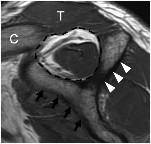
Fig. 1 MRI assessment of supraspinatus muscle volume. Supraspinatus atrophy was analyzed on most lateral image of scapular spine (arrowheads) in contact with scapular body (arrows) on oblique sagittal T1-weighted image. Imaginary line along inferior border of trapezius (T), distal clavicle (C), and scapula delineated supraspinatus fossa (dashed line). Occupation ratio of supraspinatus muscle in supraspinatus fossa was evaluated visually.
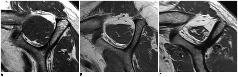
Fig. 2 Grading of supraspinatus muscle atrophy. A. Grade 1, minimal to mild atrophy of supraspinatus muscle, occupation ratio ≥ 60%. B. Grade 2, moderate atrophy of supraspinatus, occupation ratio 30-59%. C. Grade 3, severe atrophy of supraspinatus, occupation ratio < 30%.
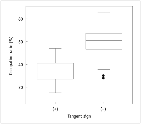
Fig. 3 Boxplot showing distribution of occupation ratio in both tangent sign positive and negative groups. Considerable overlap was observed in grade 2 muscle atrophy between two tangent sign groups. Boxes indicate first to third quartiles, each midline indicates median (second quartile), and whiskers represent maximum and minimum values within interquartile range.
Reference
-
1. Tingart MJ, Apreleva M, Lehtinen JT, Capell B, Palmer WE, Warner JJ. Magnetic resonance imaging in quantitative analysis of rotator cuff muscle volume. Clin Orthop Relat Res. 2003; (415):104–110.2. Sher JS, Uribe JW, Posada A, Murphy BJ, Zlatkin MB. Abnormal findings on magnetic resonance images of asymptomatic shoulders. J Bone Joint Surg Am. 1995; 77:10–15.3. Trudel G, Ryan SE, Rakhra K, Uhthoff HK. Extra- and intramuscular fat accumulation early after rabbit supraspinatus tendon division: depiction with CT. Radiology. 2010; 255:434–441.4. Goutallier D, Postel JM, Bernageau J, Lavau L, Voisin MC. Fatty muscle degeneration in cuff ruptures. Pre- and postoperative evaluation by CT scan. Clin Orthop Relat Res. 1994; (304):78–83.5. Thomazeau H, Rolland Y, Lucas C, Duval JM, Langlais F. Atrophy of the supraspinatus belly. Assessment by MRI in 55 patients with rotator cuff pathology. Acta Orthop Scand. 1996; 67:264–268.6. Zanetti M, Gerber C, Hodler J. Quantitative assessment of the muscles of the rotator cuff with magnetic resonance imaging. Invest Radiol. 1998; 33:163–170.7. Gladstone JN, Bishop JY, Lo IK, Flatow EL. Fatty infiltration and atrophy of the rotator cuff do not improve after rotator cuff repair and correlate with poor functional outcome. Am J Sports Med. 2007; 35:719–728.8. Goutallier D, Postel JM, Gleyze P, Leguilloux P, Van Driessche S. Influence of cuff muscle fatty degeneration on anatomic and functional outcomes after simple suture of full-thickness tears. J Shoulder Elbow Surg. 2003; 12:550–554.9. Goutallier D, Postel JM, Lavau L, Bernageau J. [Impact of fatty degeneration of the suparspinatus and infraspinatus msucles on the prognosis of surgical repair of the rotator cuff]. Rev Chir Orthop Reparatrice Appar Mot. 1999; 85:668–676.10. Liem D, Lichtenberg S, Magosch P, Habermeyer P. Magnetic resonance imaging of arthroscopic supraspinatus tendon repair. J Bone Joint Surg Am. 2007; 89:1770–1776.11. Fuchs B, Weishaupt D, Zanetti M, Hodler J, Gerber C. Fatty degeneration of the muscles of the rotator cuff: assessment by computed tomography versus magnetic resonance imaging. J Shoulder Elbow Surg. 1999; 8:599–605.12. Pfirrmann CW, Schmid MR, Zanetti M, Jost B, Gerber C, Hodler J. Assessment of fat content in supraspinatus muscle with proton MR spectroscopy in asymptomatic volunteers and patients with supraspinatus tendon lesions. Radiology. 2004; 232:709–715.13. Khoury V, Cardinal E, Brassard P. Atrophy and fatty infiltration of the supraspinatus muscle: sonography versus MRI. AJR Am J Roentgenol. 2008; 190:1105–1111.14. Sugaya H, Maeda K, Matsuki K, Moriishi J. Repair integrity and functional outcome after arthroscopic double-row rotator cuff repair. A prospective outcome study. J Bone Joint Surg Am. 2007; 89:953–960.15. Landis JR, Koch GG. The measurement of observer agreement for categorical data. Biometrics. 1977; 33:159–174.16. Resnick D, Kang HS. Internal derangements of joints: Emphasis on MR imaging. 1st ed. Philadelphia: W.B. Saunders Company;1997. p. 165–309.17. Melis B, DeFranco MJ, Chuinard C, Walch G. Natural history of fatty infiltration and atrophy of the supraspinatus muscle in rotator cuff tears. Clin Orthop Relat Res. 2010; 468:1498–1505.18. Strobel K, Hodler J, Meyer DC, Pfirrmann CW, Pirkl C, Zanetti M. Fatty atrophy of supraspinatus and infraspinatus muscles: accuracy of US. Radiology. 2005; 237:584–589.
- Full Text Links
-
- Actions
-
Cited
- CITED
-
- Close
- Share
-
- Similar articles
-
- Fatty Degeneration and Atrophy of Rotator Cuffs: Comparison of Immediate Postoperative MRI with Preoperative MRI
- Reliability of the Supraspinatus Muscle Thickness Measurement by Ultrasonography
- Quantitative Measurement of Muscle Atrophy and Fat Infiltration of the Supraspinatus Muscle Using Ultrasonography After Arthroscopic Rotator Cuff Repair
- MRI Follow-up Study After Arthroscopic Repair of Multiple Rotator Cuff Tendons
- Minimal Medial-row Tie with Suture-bridge Technique for Medium to Large Rotator Cuff Tears
