Malignant mixed tumor in the salivary gland of a cat
- Affiliations
-
- 1Department of Neurology, College of Medicine, Hanyang University, Seoul 133-792, Korea.
- 2Department of Veterinary Surgery, Faculty of Agriculture, Yamaguchi University, Yamaguchi 753-8515, Japan.
- 3Department of Veterinary Radiology, Faculty of Agriculture, Yamaguchi University, Yamaguchi 753-8515, Japan. nakaichi@yamaguchi-u.ac.jp
- KMID: 1718588
- DOI: http://doi.org/10.4142/jvs.2008.9.3.331
Abstract
- The presence of a malignant mixed tumor, also known as a carcinosarcoma, in the salivary gland is very rare. Such mors, which are typically aggressive, are characterized by the presence of carcinomatous and sarcomatous components. 9-year-old neutered female domestic short-haired cat presented with swelling in the right mandibular lesion that had rapidly enlarged over the previous 3 weeks. Physical examination revealed a large, fluctuated and painless subcutaneous swelling that was associated with a firm mass. Radiographs of the head revealed a soft-tissue density that involved faint circular calcific opacity. Contrast-enhanced computed tomography revealed that the peripheral capsulated cystic area had a contrast enhanced region without bone lysis. The cat received a total excision of the mass and postoperative radiotherapy. Histopathological analysis of the mass revealed that it was a malignant mixed tumor. Metastasis to the lung was discovered 7 weeks later, at which time treatment was stopped.
MeSH Terms
Figure
-
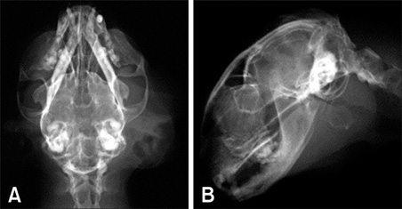
Fig. 1 Radiographic images of the head showing a large tissue-dense mass (A) with faint circular calcific opacity (B).

Fig. 2 Post-contrast CT images showing enlargement of the right submandibular gland with a peripheral capsulated cystic area (A: tympanic bulla level), and contrast enhanced region (B: occipital bone level, C: first cervical vertebra level) that was believed to be a tumor.
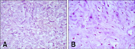
Fig. 3 Adenocarcinomatous (A) and chondrosarcomatous (B) elements were observed in the specimen. H&E stain. ×400.
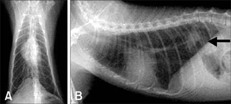
Fig. 4 Thoracic radiographs showed a diffuse increase in the distribution of pulmonary interstitial densities and a poorly circumscribed-nodular mass in the caudodorsal lung lobe (arrow), suggesting lung metastasis of the tumor (A: ventrodorsal view, B: lateral view).
Reference
-
1. Carberry CA, Flanders JA, Harvey HJ, Ryan AM. Salivary gland tumors in dogs and cats: a literature and case review. J Am Anim Hosp Assoc. 1988. 24:561–567.2. Carpenter JL, Bernstein M. Malignant mixed (pleomorphic) mandibular salivary gland tumors in a cat. J Am Anim Hosp Assoc. 1991. 27:581–583.3. Ellis GL, Gnepp DR. Gnepp DR, editor. Unusual salivary gland tumors. Pathology of the Head and Neck. 1988. New York: Churchill Livingstone;623–627.4. Galli J, Parrilla C, Corina L, Ricci R, Paludetti G. Carcinosarcoma of the submandibular salivary gland: clinical case and review of the literature. J Otolaryngol. 2005. 34:66–69.
Article5. Gnepp DR. Malignant mixed tumors of the salivary glands: a review. Pathol Annu. 1993. 28:279–328.6. Hammer A, Getzy D, Ogilvie G, Upton M, Klausner J, Kisseberth WC. Salivary gland neoplasia in the dog and cat: survival times and prognostic factors. J Am Anim Hosp Assoc. 2001. 37:478–482.
Article7. Head KW, Cullen JM, Dubielzig RR, Else RW, Misdorp W, Patnaik AK, Tateyama S, van der Gaag I. Histologic Classification of Tumors of the Alimentary System of Domestic Animals. 2003. Armed Forces Institute of Pathology: Washington DC;58–72. 2nd series.8. Horky JK, Chaloupka JC, Putman CM, Roth TC, Weaver EM, Sasaki CT. True malignant mixed tumor (carcinosarcoma) of tonsillar minor salivary gland origin: diagnostic imaging and endovascular therapeutic embolization. AJNR Am J Neuroradiol. 1997. 18:1944–1948.9. King AD, Ahuja AT, To EW, Chan EC, Allen PW. Carcinosarcoma of the parotid gland: ultrasound and computed tomography findings. Australas Radiol. 1999. 43:520–522.
Article10. Kwon MY, Gu M. True malignant mixed tumor (carcinosarcoma) of parotid gland with unusual mesenchymal component. Arch Pathol Lab Med. 2001. 125:812–815.
Article11. Lai PH, Chang JM, Hou YY, Chu ST, Lin SL, Yang CF. Carcinosarcoma of the salivary gland on CT. AJNR Am J Neuroradiol. 1995. 16:1733–1735.12. Magnano M, Gervasio CF, Cravero L, Machetta G, Lerda W, Beltramo G, Orecchia R, Ragona R, Bussi M. Treatment of malignant neoplasms of the parotid gland. Otolaryngol Head Neck Surg. 1999. 121:627–632.
Article13. Owaki S, Kitano H, Hanada M, Asada Y, Sugihara H, Moritani S, Banba M. Carcinosarcoma of the submandibular gland: an autopsy case. Auris Nasus Larynx. 2003. 30:439–442.
Article14. Stephen J, Batsakis JG, Luna MA, von der Heyden U, Byers RM. True malignant mixed tumors (carcinosarcoma) of salivary glands. Oral Surg Oral Med Oral Pathol. 1986. 61:597–602.
Article15. Tortoledo ME, Luna MA, Batsakis JG. Carcinomas ex pleomorphic adenoma and malignant mixed tumors. Histomorphologic indexes. Arch Otolaryngol. 1984. 110:172–176.
Article16. Wells GAH, Robinson M. Mixed tumor of salivary gland showing histological evidence of malignancy in a cat. J Comp Pathol. 1975. 85:77–85.
Article17. Withrow SJ. Withrow SJ, MacEwen EG, editors. Cancer of the salivary glands. Small Animal Clinical Oncology. 1996. 2nd ed. Philadelphia: Saunders;240–241.
- Full Text Links
-
- Actions
-
Cited
- CITED
-
- Close
- Share
-
- Similar articles
-
- A Case of Giant Carcinosarcoma of the Parotid Gland
- A Case of Multiple Cutaneous Metastases from Metastasizing PleomorphicAdenoma of the Submandibular Gland
- A Case of Salivary Duct Carcinoma Ex Pleomorphic Adenoma of Parotid Gland
- A Case of Mucoepidermoid Carcinoma Mixed with Osteosarcoma of the Parotid Gland Extending to the Parapharyngeal Space
- Carcinoma ex mixed tumor arising in the parotid gland
