A Case of Pancreatic Pseudocyst with a Large Subcapsular Splenic Hematoma Treated Successfully by Ultrasonography-guided Percutaneous Drainage
- Affiliations
-
- 1Department of Internal Medicine, Chonnam National University Medical School, Gwangju, Korea. choisk@chonnam.ac.kr
- KMID: 1718408
- DOI: http://doi.org/10.4166/kjg.2011.57.4.258
Abstract
- A subcapsular splenic hematoma is a very rare hemorrhagic complication of pancreatitis. We report here on a case of pseudocyst with a large subcapsular splenic hematoma in a 43-year-old man who presented with severe left flank pain for one week. Despite the initial conservative treatment consisting of pain control, bowel rest, intravenous fluids and antibiotics, the pain was not relieved. An abdominal computed tomography (CT) was performed, and it showed a pseudocyst that was increasing in size with a large subcapsular splenic hematoma measuring 6x13 cm compared to the images at admission. Ultrasonography (US)-guided percutaneous drainage was performed without any complications, and splenectomy was avoided. After the discharge, the patient remained asymptomatic for eight months. We suggest that percutaneous drainage of a large subcapsular hematoma complicating pancreatitis might be a useful treatment option in selected patients.
MeSH Terms
Figure
-
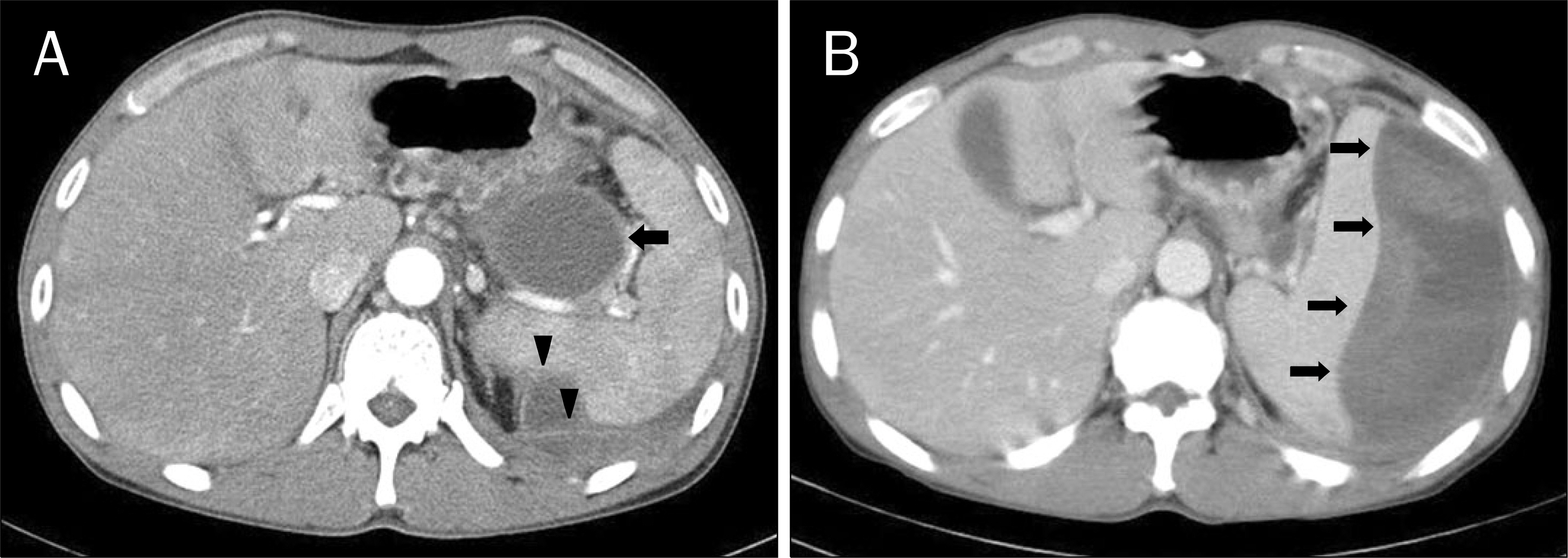
Fig. 1. Computed tomographic (CT) scan images during the second episode of pancreatitis and in the emergency room. (A) Variable sized multiple pseudocysts in the tail of the pancreas compressed the adjacent structures including splenic vein, gastric fundus (arrow), and splenic parenchyma (arrow heads). (B) A CT scan showed increased size of the pseudocyst with a large subcapsular splenic hematoma (arrows) measuring 5×12 cm in the tail of pancreas.
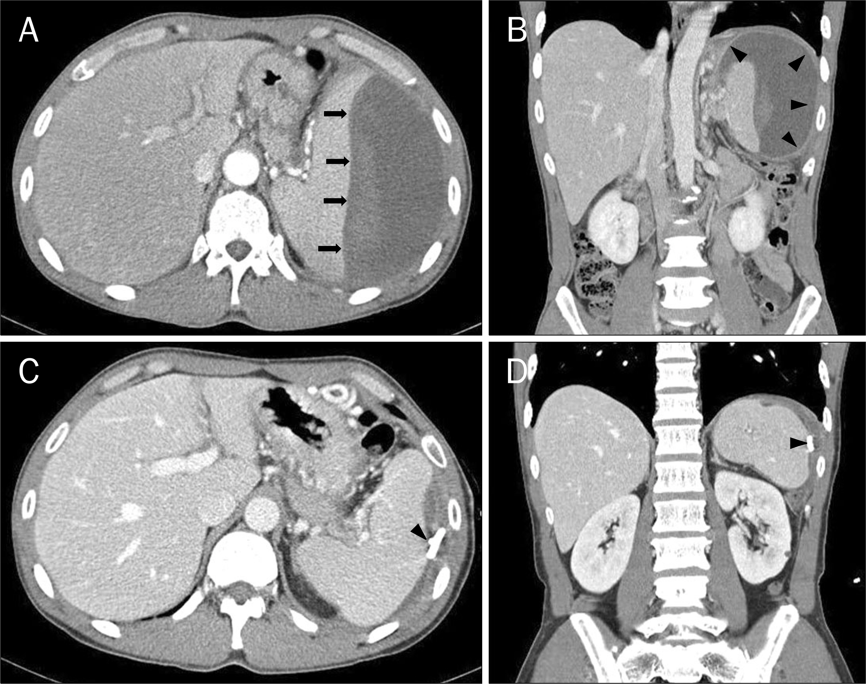
Fig. 2. CT scan images on hospital day 6 and 3 weeks after US-guided drainage. (A and B) increased size (approximately 6×13 cm) of the psedocyst with subcapsular splenic hematoma is shown (arrows and arrow heads). (C and D) CT images 3 weeks after US-guided percutaneous drainage, the size of the pseudocyst with subcapsular splenic hematoma was markedly decreased. A drainage catheter is shown (arrow head).
Reference
-
References
1. Habashi S, Draganov PV. Pancreatic pseudocyst. World J Gastroenterol. 2009; 15:38–47.
Article2. Kuramitsu T, Komatsu M, Ono T, et al. Ruptured subcapsular giant hematoma of the spleen as a complication of chronic pancreatitis. Intern Med. 1995; 34:564–568.
Article3. Vyborny CJ, Merrill TN, Reda J, Geurkink RE, Smith SJ. Subacute subcapsular hematoma of the spleen complicating pancreatitis: successful percutaneous drainage. Radiology. 1988; 169:161–162.
Article4. Malka D, Hammel P, Lévy P, et al. Splenic complications in chronic pancreatitis: prevalence and risk factors in a medical-surgical series of 500 patients. Br J Surg. 1998; 85:1645–1649.
Article5. Siu TL. Percutaneous drainage of spontaneous subcapsular haematoma of the spleen complicating chronic pancreatitis. Surgeon. 2004; 2:52–55.
Article6. Patel VG, Eltayeb OM, Zakaria M, Fortson JK, Weaver WL. Spontaneous subcapsular splenic hematoma: a rare complication of pancreatitis. Am Surg. 2005; 71:1066–1069.
Article7. Tseng CW, Chen CC, Chiang JH, Chang FY, Lin HC, Lee SD. Percutaneous drainage of large subcapsular hematoma of the spleen complicating acute pancreatitis. J Chin Med Assoc. 2008; 71:92–95.
Article8. Lankisch PG. The spleen in inflammatory pancreatic disease. Gastroenterology. 1990; 98:509–516.
Article9. Greenstein A, DeMaio EF, Nabseth DC. Acute hemorrhage associated with pancreatic pseudocysts. Surgery. 1971; 69:56–62.10. Warshaw AL, Chesney TM, Evans GW, McCarthy HF. Intrasplenic dissection by pancreatic pseudocysts. N Engl J Med. 1972; 287:72–75.
Article11. Sitzmann JV, Imbembo AL. Splenic complications of a pancreatic pseudocyst. Am J Surg. 1984; 147:191–196.
Article12. Heider R, Behrns KE. Pancreatic pseudocysts complicated by splenic parenchymal involvement: results of operative and percutaneous management. Pancreas. 2001; 23:20–25.
Article13. Agnifili A, Gianfelice F, Gola P, et al. Subcapsular splenic hematoma complicating chronic relapsing pancreatitis. Case report. Eur J Surg. 1991; 157:63–65.
- Full Text Links
-
- Actions
-
Cited
- CITED
-
- Close
- Share
-
- Similar articles
-
- Large Subcapsular Pseudocyst of Spleen Complicated by Chronic Pancreatitis
- Percutaneous drainage of pancreatic abscess and pseudocyst
- Percutaneous Catheter Drainage of Pancreatic Pseudocyst through the Kidney: A case Report
- A Case of Pancreatic Pseudocyst in the Left Hepatic Lobe
- A Case of Splenic Pseudocyst Complicated by Acute Pancreatitis
