A Case of Clonorchiasis Presenting as Common Bile Duct Mass
- Affiliations
-
- 1Department of Internal Medicine, Catholic University of Daegu School of Medicine, Daegu, Korea. hgkim@cu.ac.kr
- KMID: 1718355
- DOI: http://doi.org/10.4166/kjg.2010.56.4.211
Abstract
- No abstract available.
MeSH Terms
-
Aged, 80 and over
Animals
Anthelmintics/therapeutic use
Bile Duct Neoplasms/diagnosis/surgery/ultrasonography
Cholangiopancreatography, Endoscopic Retrograde
Clonorchiasis/*diagnosis/drug therapy/surgery
Clonorchis sinensis/isolation & purification
Common Bile Duct/ultrasonography
Humans
Male
Praziquantel/therapeutic use
Tomography, X-Ray Computed
Figure
-

Fig. 1. Contrast-enhanced CT finding of the patient. There were gallbladder distension and wall thickening, and dilated common bile duct.

Fig. 2. Endoscopic retrograde cholangiopancreatographic finding. Major papilla looked normal in endoscopic view.
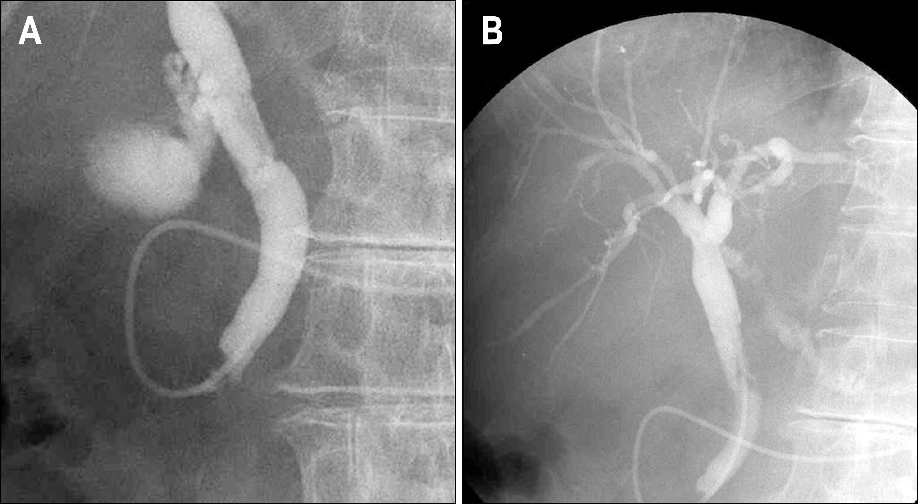
Fig. 3. Endoscopic retrograde cholangiopancreatographic findings. (A) A filling defect was noted at the distal common bile duct. (B) Threre was diffuse mild dilatation at peripheral intrahepatic bile ducts.
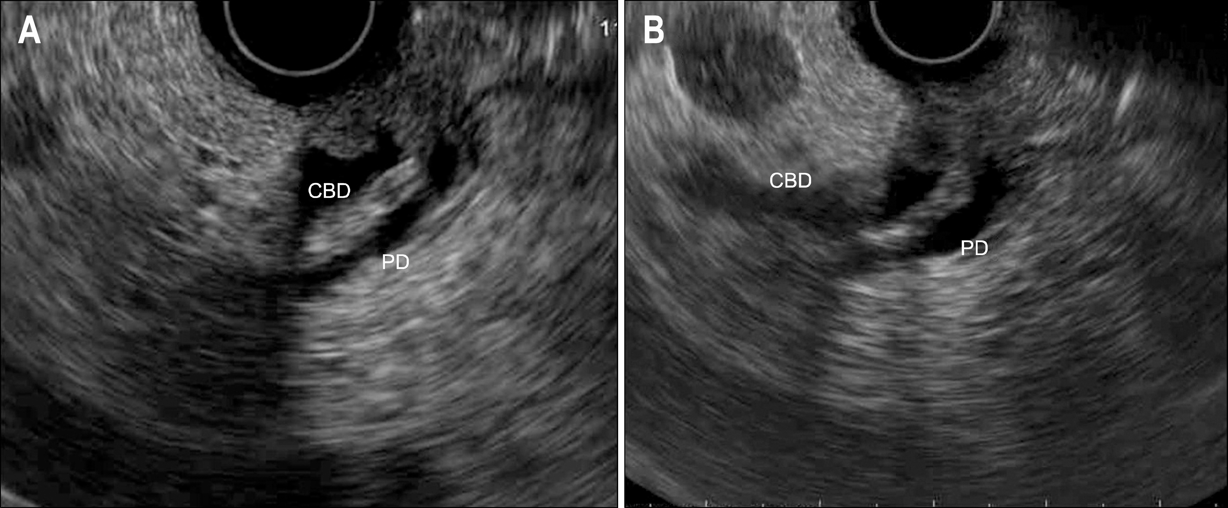
Fig. 4. Endoscopic ultrasound findings. There was a small papillary mass in the distal common bile duct. CBD, common bile duct; PD, pancreatic duct.

Fig. 5. Endoscopic view after second endoscopic retrograde cholangiopancreatography. (A) After endoscopic sphincterotomy, a part of fluke was seen in the ampulla. (B) The flukes were extracted into the duodenum through the ampulla with balloon basket.
Cited by 1 articles
-
A Case of Clonorchiasis with Focal Intrahepatic Duct Dilatation Mimicking an Intrahepatic Cholangiocarcinoma
Bong Gap Kim, Dae Hwan Kang, Cheol Woong Choi, Hyung Wook Kim, Jae Hyung Lee, Suk Hun Kim, Hye Ju Yeo, Soo Yong Lee
Clin Endosc. 2011;44(1):55-58. doi: 10.5946/ce.2011.44.1.55.
Reference
-
1. Kim HG, Han J, Kim MH, et al. Prevalence of clonorchiasis in patients with gastrointestinal disease: A Korean nationwide multicenter survey. World J Gastroenterol. 2009; 15:86–94.
Article2. Joo CY, Chung MS, Kim SJ, Kang CM. Changing patterns of clonorchis sinensis infections in Kyongbuk, Korea. Korean J Parasitol. 1997; 35:155–164.
Article3. Kim YH. Extrahepatic cholangiocarcinoma associated with clonorchiasis: CT evaluation. Abdom Imaging. 2003; 28:68–71.
Article4. Lim JH. Radiologic findings of clonorchiasis. AJR Am J Roentgenol. 1990; 155:1001–1008.
Article5. Bhargava DK. Endoscopy and biliary parasites. Gastrointest Endosc Clin N Am. 1996; 6:139–152.
Article6. Park YH, Choi SK. A clinical review on biliary clonorchiasis. Korean J Gastroenterol. 1986; 18:145–152.7. Song HY, Rhee KS, Lee ST, Kim DK, Ahn DS. Clinical features in clonorchiasis. Korean J Gastroenterol. 1995; 27:64–71.8. Kang DH, Choi SH, Chun KJ, et al. ERCP findings in hepatic clonorchiasis. Korean J Gastrointest Endosc. 1993; 13:121–126.
- Full Text Links
-
- Actions
-
Cited
- CITED
-
- Close
- Share
-
- Similar articles
-
- Radiological findings of clonorchiasis
- A Case of Villous Adenomas in the Common Bile Duct and Cystic Duct
- A Case of Clonorchiasis Presenting as Common Hepatic Duct Mass
- Too many ducts sign: a characteristic cholangiographic finding of clonorchiasis?
- Radiologic imaging of bile duct changes by clonorchiasis
