Magnetic Resonance Arthrographic Dissection of Posterolateral Corner of the Knee: Revealing the Meniscofibular Ligament
- Affiliations
-
- 1Department of Radiology, Research Institute of Radiological Science, Medical Convergence Research Institute, and Severance Biomedical Science Institute, Yonsei University College of Medicine, Seoul, Korea. jss@yuhs.ac
- 2Department of Orthopedic Surgery and Arthroscopic Surgery Unit, Yonsei University College of Medicine, Seoul, Korea.
- KMID: 1716884
- DOI: http://doi.org/10.3349/ymj.2012.53.4.820
Abstract
- PURPOSE
To evaluate meniscofibular ligament (MFibL) at the posterolateral corner of the knee joint on the magnetic resonance arthrography (MRA) with 70degrees knee flexion.
MATERIALS AND METHODS
The MRA of the knee joint was performed at 70degrees knee flexion. Eighteen patients (19 knee joints) underwent scanning of sagittal, coronal, and axial fat-suppressed T1 weighted images (T1FS), and coronal fat-suppressed T2 weighted images. Sagittal three-dimensional (3D) gradient echo (GRE) images were also obtained. Retrospective review of 19 knee MRA studies was independently performed by two musculoskeletal radiologists. The statistical significance was proved by chi-square test.
RESULTS
The MFibL ligament was optimally demonstrated on the far lateral sagittal 3D GRE and T1FS MRA images. The MFibL appeared as a curvilinear or straight hypointense band of variable thickness, extended from the posterolateral meniscus to upper anteromedial aspect of the fibular head. The MFibL was demonstrated with scale 2 (more than a half length of the ligament) by both reviewers in 73.68% (n=14/19) of the knee 3D GRE images and 89.47% (n=17/19) of the knee T1FS images. The visualization on T1FS and that on GRE were not statistically different from each other (p>0.05). The interobserver agreements were significantly good on both 3D GRE and T1FS images in detecting the ligament (kappa values, 0.642 and 0.683, respectively).
CONCLUSION
The MFibL is well visualized on the far lateral sagittal MRA at 70degrees knee flexion, which could potentially be useful in recognizing structures in the posterolateral corner of the knee, including the MFibL.
MeSH Terms
Figure
-
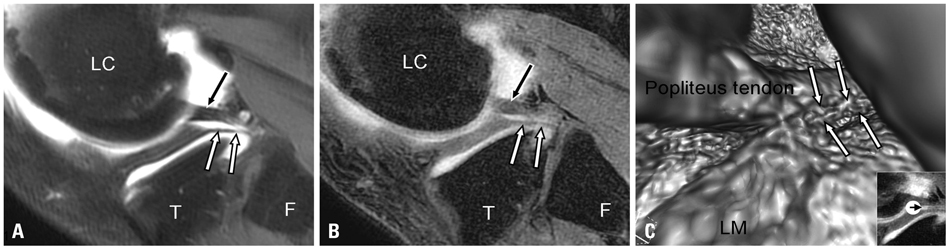
Fig. 1 MR arthrographic appearance of the meniscofibular ligament (MFibL). (A and B) A sagittal fat suppressed T1 weighted image of MR arthrography (A) and a corresponding sagittal 3D gradient echo MR arthrography (B) demonstrate the MFibL as a linear hypointense structure (white arrows) extending between the apex of the fibular head and the posterior third of the lateral meniscus. The popliteus tendon is located posterior to MFibL (black arrow). (C) Virtual MR arthroscopy reveals the relationship between the popliteus tendon and the MFibL (white arrows). LC, lateral condyle of femur; T, tibia; F, fibula; LM, lateral meniscus; MR, magnetic resonance; 3D, three-dimensional.
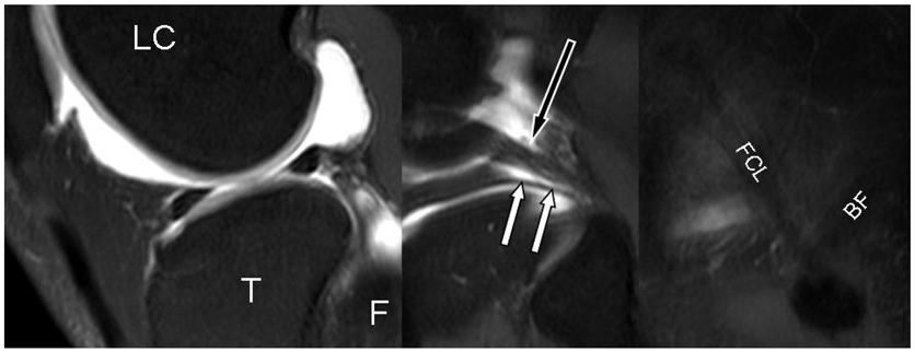
Fig. 2 MR arthrographic appearance of meniscofibular ligament (MFibL). Continuous sagittal fat suppressed T1 weighted images of knee MR arthrography demonstrate the MFibL as a linear, hypointense structure (white arrows), extending between the apex of the fibular head and the posterior third of the lateral meniscus. The popliteus tendon is located posterior to MFibL (black arrow). LC, lateral condyle of femur, T, tibia; F, fibula; FCL, fibular collateral ligament; BF, biceps femoris tendon; MR, magnetic resonance.
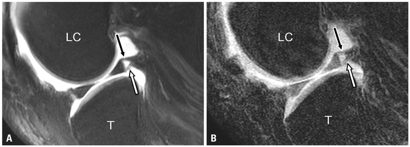
Fig. 3 Posterosuperior and posteroinferior popliteomeniscal fascicules. A sagittal fat suppressed T1 weighted image of MR arthrography (A) and a corresponding sagittal 3D gradient echo MR arthrography (B) demonstrate the posteosuperior (black arrow) and posteroinferior (white arrow) popliteomeniscal fascicles. LC, lateral condyle of femur; T, tibia; MR, magnetic resonance; 3D, three-dimensional.

Fig. 4 The difference between fat suppressed T1 weighted image and 3D gradient echo image. A sagittal fat suppressed T1 weighted image of MR arthrography (A) demonstrates the MFibL as a linear hypointense structure (white arrows). The popliteus tendon is located posterior to the MFibL (black arrow). However, the MFibL can partially be delineated on the corresponding sagittal 3D gradient echo MR arthrography (B). LC, lateral condyle of femur; T, tibia; F, fibula; MFibL, meniscofibular ligament; MR, magnetic resonance; 3D, three-dimensional.
Reference
-
1. Covey DC. Injuries of the posterolateral corner of the knee. J Bone Joint Surg Am. 2001. 83-A:106–118.
Article2. Bozkurt M, Elhan A, Tekdemir I, Tönük E. An anatomical study of the meniscofibular ligament. Knee Surg Sports Traumatol Arthrosc. 2004. 12:429–433.
Article3. Standring S. Gray's anatomy: The Anatomical Basis of Clinical Practice. 2005. 39th ed. Elsevier Churchill-Livingstone;1477.4. Obaid H, Gartner L, Haydar AA, Briggs TW, Saifuddin A. The meniscofibular ligament: an MRI study. Eur J Radiol. 2010. 73:159–161.
Article5. Munshi M, Pretterklieber ML, Kwak S, Antonio GE, Trudell DJ, Resnick D. MR imaging, MR arthrography, and specimen correlation of the posterolateral corner of the knee: an anatomic study. AJR Am J Roentgenol. 2003. 180:1095–1101.
Article6. Krudwig WK, Witzel U, Ullrich K. Posterolateral aspect and stability of the knee joint. II. Posterolateral instability and effect of isolated and combined posterolateral reconstruction on knee stability: a biomechanical study. Knee Surg Sports Traumatol Arthrosc. 2002. 10:91–95.
Article7. Ogden JA. The anatomy and function of the proximal tibiofibular joint. Clin Orthop Relat Res. 1974. 186–191.8. Bozkurt M, Yilmaz E, Atlihan D, Tekdemir I, Havitçioğlu H, Günal I. The proximal tibiofibular joint: an anatomic study. Clin Orthop Relat Res. 2003. 136–140.9. Peduto AJ, Nguyen A, Trudell DJ, Resnick DL. Popliteomeniscal fascicles: anatomic considerations using MR arthrography in cadavers. AJR Am J Roentgenol. 2008. 190:442–448.
Article10. Terry GC, LaPrade RF. The posterolateral aspect of the knee. Anatomy and surgical approach. Am J Sports Med. 1996. 24:732–739.11. Ullrich K, Krudwig WK, Witzel U. Posterolateral aspect and stability of the knee joint. I. Anatomy and function of the popliteus muscle-tendon unit: an anatomical and biomechanical study. Knee Surg Sports Traumatol Arthrosc. 2002. 10:86–90.
Article
- Full Text Links
-
- Actions
-
Cited
- CITED
-
- Close
- Share
-
- Similar articles
-
- Treatment of Posterolateral Rotatory Instability of the Knee
- Common Peroneal Nerve Palsy Associated with Multiple Knee Ligament Injury
- Posterior Cruciate Ligament Reconstruction and Posterolateral Corner Injury
- Anatomic Reconstructive Surgery of the Posterolateral Instability of the Knee Using Split Acilles Allograft
- Magnetic Resonance Imaging for the Evaluation of Acute Posterolateral Complex Injuries of the Knee
