Yonsei Med J.
2012 Jul;53(4):680-684. 10.3349/ymj.2012.53.4.680.
Comparison between Measured and Calculated Length of Side Branch Ostium in Coronary Bifurcation Lesions with Intravascular Ultrasound
- Affiliations
-
- 1Division of Cardiology, Gumi CHA University Medical Center, Gumi, Korea.
- 2Division of Cardiology, Severance Cardiovascular Hospital, Yonsei University College of Medicine, Seoul, Korea. mkhong61@yuhs.ac
- 3Division of Cardiology, Severance Biomedical Science Institute, Yonsei University College of Medicine, Seoul, Korea.
- KMID: 1716863
- DOI: http://doi.org/10.3349/ymj.2012.53.4.680
Abstract
- PURPOSE
Accurate evaluation of side branch (SB) ostium could be critical to the treatment of bifurcation lesions. We compared measured and calculated values of side branch ostial length (SBOL) in coronary bifurcation lesions with intravascular ultrasound (IVUS).
MATERIALS AND METHODS
Pre-intervention and post-intervention IVUS was performed in 113 patients who underwent stent implantation of bifurcation lesions. For the IVUS longitudinal reconstruction of the bifurcation lesions, SBOL, SB diameter, and the angle between the distal portion of the main vessel (MV) and SB were directly measured. In addition, SBOL was calculated as: SB diameter/sin (angle between distal MV and SB). The relationship between measured and calculated SBOL was then evaluated.
RESULTS
The angled between the distal MV and SB were 57.3+/-12.4degrees at pre-intervention and 59.4+/-12.6degrees at post-intervention. The mean measured and calculated SBOL values were 2.91+/-0.86 mm and 3.06+/-0.77 mm at pre-intervention and 2.79+/-0.82 mm and 2.92+/-0.69 mm at post-intervention, respectively. Differences between measured and calculated SBOL were 0.15+/-0.44 mm at pre-intervention and 0.13+/-0.41 mm at post-intervention. We found that calculated SBOL was correlated with measured SBOL (pre-intervention r=0.863, p<0.001; post-intervention r=0.868, p<0.001).
CONCLUSION
There was a good correlation between measured and calculated SBOLs of the bifurcation lesions in IVUS longitudinal reconstruction. SBOL in the bifurcation lesions can therefore be estimated using the SB diameter and the angle between distal MV and SB.
Keyword
MeSH Terms
Figure
-

Fig. 1 Intravascular ultrasound (IVUS) measurements in this study are shown in a schematic diagram (A) and an actual IVUS image (B); side branch diameter (a), angle between distal main vessel and side branch (b), and side branch ostium length (c).
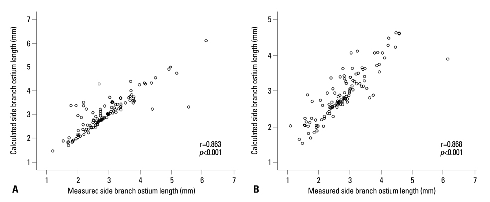
Fig. 2 The correlation between the measured and calculated values of the side branch ostium length are shown; pre-intervention r=0.863, p<0.001 (A) and post-intervention r=0.868, p<0.001 (B).
Reference
-
1. Latib A, Colombo A, Sangiorgi GM. Bifurcation stenting: current strategies and new devices. Heart. 2009. 95:495–504.
Article2. Aliabadi D, Tilli FV, Bowers TR, Benzuly KH, Safian RD, Goldstein JA, et al. Incidence and angiographic predictors of side branch occlusion following high-pressure intracoronary stenting. Am J Cardiol. 1997. 80:994–997.
Article3. Fischman DL, Savage MP, Leon MB, Schatz RA, Ellis S, Cleman MW, et al. Fate of lesion-related side branches after coronary artery stenting. J Am Coll Cardiol. 1993. 22:1641–1646.
Article4. Pan M, Medina A, Suárez de Lezo J, Romero M, Melián F, Pavlovic D, et al. Follow-up patency of side branches covered by intracoronary Palmaz-Schatz stent. Am Heart J. 1995. 129:436–440.
Article5. Hoye A, Iakovou I, Ge L, van Mieghem CA, Ong AT, Cosgrave J, et al. Long-term outcomes after stenting of bifurcation lesions with the "crush" technique: predictors of an adverse outcome. J Am Coll Cardiol. 2006. 47:1949–1958.
Article6. Moussa I, Costa RA, Leon MB, Lansky AJ, Lasic Z, Cristea E, et al. A prospective registry to evaluate sirolimus-eluting stents implanted at coronary bifurcation lesions using the "crush technique". Am J Cardiol. 2006. 97:1317–1321.
Article7. Latib A, Colombo A. Bifurcation disease: what do we know, what should we do? JACC Cardiovasc Interv. 2008. 1:218–226.8. Costa RA, Mintz GS, Carlier SG, Lansky AJ, Moussa I, Fujii K, et al. Bifurcation coronary lesions treated with the "crush" technique: an intravascular ultrasound analysis. J Am Coll Cardiol. 2005. 46:599–605.9. Mintz GS, Weissman NJ. Intravascular ultrasound in the drug-eluting stent era. J Am Coll Cardiol. 2006. 48:421–429.
Article10. Park SJ, Kim YH, Park DW, Lee SW, Kim WJ, Suh J, et al. Impact of intravascular ultrasound guidance on long-term mortality in stenting for unprotected left main coronary artery stenosis. Circ Cardiovasc Interv. 2009. 2:167–177.
Article11. Kim JS, Hong MK, Ko YG, Choi D, Yoon JH, Choi SH, et al. Impact of intravascular ultrasound guidance on long-term clinical outcomes in patients treated with drug-eluting stent for bifurcation lesions: data from a Korean multicenter bifurcation registry. Am Heart J. 2011. 161:180–187.
Article12. Mintz GS, Nissen SE, Anderson WD, Bailey SR, Erbel R, Fitzgerald PJ, et al. American College of Cardiology Clinical Expert Consensus Document on Standards for Acquisition, Measurement and Reporting of Intravascular Ultrasound Studies (IVUS). A report of the American College of Cardiology Task Force on Clinical Expert Consensus Documents. J Am Coll Cardiol. 2001. 37:1478–1492.
Article13. Lima T, Alves C, Funayama CA. Proposal for a trigonometric method to evaluate the abduction angle of the lower limbs in neonates. J Child Neurol. 2008. 23:1451–1454.
Article14. Al Suwaidi J, Yeh W, Cohen HA, Detre KM, Williams DO, Holmes DR Jr. Immediate and one-year outcome in patients with coronary bifurcation lesions in the modern era (NHLBI dynamic registry). Am J Cardiol. 2001. 87:1139–1144.
Article15. Sano K, Mintz GS, Carlier SG, de Ribamar Costa J Jr, Qian J, Missel E, et al. Assessing intermediate left main coronary lesions using intravascular ultrasound. Am Heart J. 2007. 154:983–988.
Article16. Furukawa E, Hibi K, Kosuge M, Nakatogawa T, Toda N, Takamura T, et al. Intravascular ultrasound predictors of side branch occlusion in bifurcation lesions after percutaneous coronary intervention. Circ J. 2005. 69:325–330.
Article
- Full Text Links
-
- Actions
-
Cited
- CITED
-
- Close
- Share
-
- Similar articles
-
- Consecutive Jailed- and Kissing-Corsair Technique: Side Branch Protection and Dilation during Stent Implantation
- Predictors of Side Branch Occlusion Immediately after Coronary Stenting: an Intravascular Ultrasound Study
- A Case of Coronary Pseudostenosis, Diagnosed by Intravascular Ultrasound
- Comparison of Angiographic Outcomes of Side Branch Ostium at Bifurcation Coronary Lesion between Two-stent and One-stent Techniques
- Percutaneous Treatment of an Injured Coronary Stent Using the Looping Wire Technique
