C-1 Root Schwannoma with Aggressive Lateral Mass Invasion
- Affiliations
-
- 1Department of Neurosurgery, Medical College, Korea University, Seoul, Korea.
- 2Department of Diagnostic Pathology, Medical College, Korea University, Seoul, Korea.
- KMID: 1716531
- DOI: http://doi.org/10.3349/ymj.2005.46.4.575
Abstract
- Schwannomas are relatively common, benign tumors that are thought to arise from the nerve sheath cell. Schwannomas of the C1 root are extremely rare and seldom invade lateral masses because they gradually increase in size and can extend through the wide space behind the lateral mass instead of the intervertebral foramen. We present here an unusual case of a benign schwannoma that aggressively invaded the lateral mass of C-1.
MeSH Terms
Figure
-
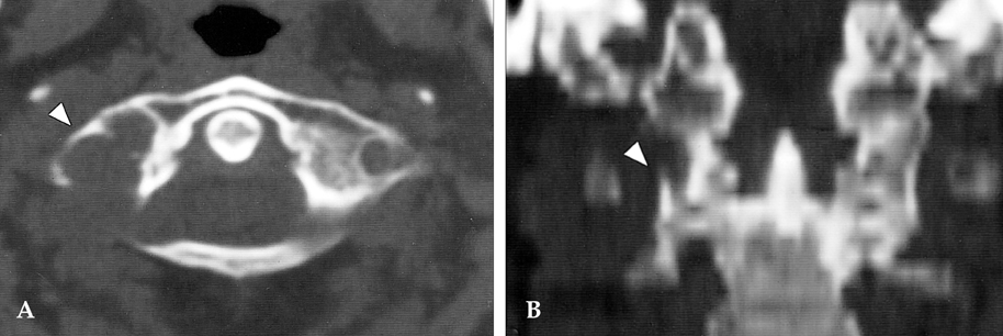
Fig. 1 A and B: Pre-operative CT scan with bone setting reveals extensive erosion with marginal sclerosis in the right transverse process and superior articular facet of C-1 (white triangle).
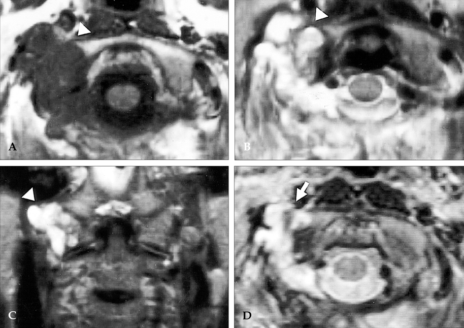
Fig. 2 (A, B) Axial T1-weighted MRI scan reveals a multilobulated mass involving the lateral mass of C-1 and enhancing moderately with indefinite tumor margin (white triangle). (C) Coronal T2-weighted MRI scan reveals mass in the right articular facet of C-1 spine (white triangle). (D) After 1 year, MRI scan demonstrates the remnant of the tumor after radiotherapy (white arrow).
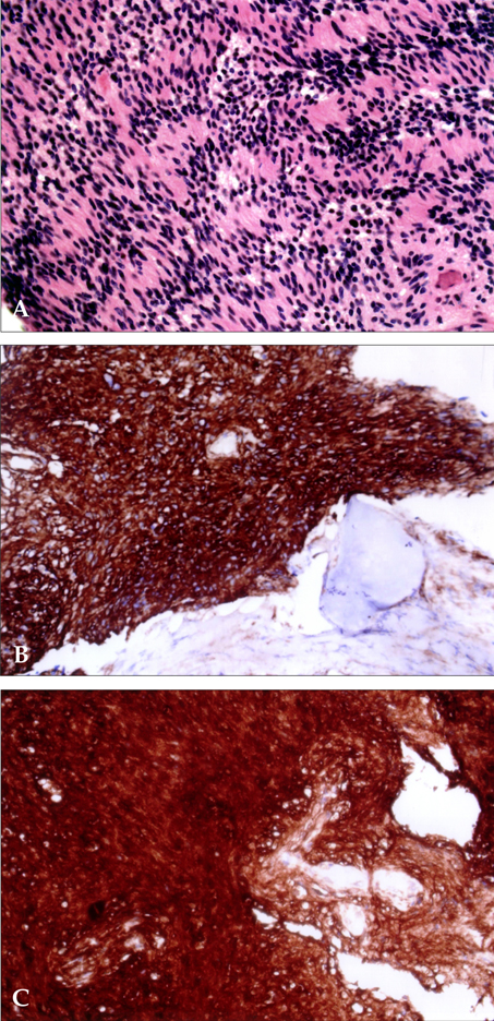
Fig. 3 Histological examination reveals a schwannoma composed of interwoven bundles of spindle-shaped, fibrillated cells (hematoxylin and eosin, 200×, A) and immunopositivity for vimentin(200×, B) and S-100 (200×, C).
Reference
-
1. Kalavakonda C, Sekhar LN, Jones RV, Rehaman AB. Intermittent vertebral artery compression caused by C1-root schwannoma: case report. Neurol Res. 2000. 22:679–684.2. Lot G, George B. Cervical neuromas with extradural components: surgical management in a series of 57 patients. Neurosurgery. 1997. 41:813–820.3. Barnowsky L, Dalal R. Extradural schwannoma manifested as an expansile vertebral lesion. AJR Am J Roentgenol. 1992. 159:1352–1353.4. George B, Lot G. Neurinomas of the first two cervical nerve roots: a series of 42 cases. J Neurosurg. 1995. 82:917–923.5. Guidetti B, Spallone A. Benign extramedullary tumors of the foramen magnum. Adv Tech Stand Neurosurg. 1988. 16:83–120.6. Inaoka T, Takahashi K, Hanaoka H, Aburano R, Tokusashi Y, Matsuno T, et al. Paravertebral neurinoma associated with aggressive intravertebral extension. Skeletal Radiol. 2001. 30:286–289.7. Kumar R, Guinto FC Jr, Madewell JE, David R, Shirkhoda A. Expansile bone lesions of the vertebra. Radiographics. 1988. 8:749–769.
- Full Text Links
-
- Actions
-
Cited
- CITED
-
- Close
- Share
-
- Similar articles
-
- Minimally Invasive Oblique Retroperitoneal Approach for Extraforaminal Lumbar Schwannoma: Technical Challenges and Literature Review
- Schwannoma on the Lateral Side of the Proximal Thigh
- A Case of Large Foramen Magnum Schwannoma
- Schwannoma Diagnosed with Duplex Ultrasonography
- Vestibular Schwannoma Atypically Invading Temporal Bone
