Yonsei Med J.
2006 Feb;47(1):148-151. 10.3349/ymj.2006.47.1.148.
Deep Femoral Artery Aneurysm Presenting as Lower Limb Swelling: A Case Report
- Affiliations
-
- 1Department of Surgery, University of Ulsan College of Medicine, Gangneung Asan Hospital, Gangneung, Korea. ypcho@gnah.co.kr
- 2Department of Diagnostic Radiology, University of Ulsan College of Medicine, Gangneung Asan Hospital, Gangneung, Korea.
- 3Department of Surgery, University of Ulsan College of Medicine, Seoul Asan Hospital, Seoul, Korea.
- KMID: 1715887
- DOI: http://doi.org/10.3349/ymj.2006.47.1.148
Abstract
- We report a rare case of lower limb swelling due to compression of the superficial femoral vein by a solitary deep femoral artery aneurysm. The patient was a 58-year-old man presenting with acute swelling of the right lower limb caused by deep venous thrombosis. A multi-detector computed tomographic scan (CT) confirmed the diagnosis of a deep femoral artery aneurysm and revealed no evidence of aneurysms or occlusive lesions in the other arteries. Aneurysmectomy and ligation of the deep femoral artery were successfully performed. Preoperative multi-detector CT scanning is a valuable, non-invasive diagnostic tool to delineate not only the state of the aneurysm, but also that of the distal arterial tree.
Keyword
MeSH Terms
Figure
-
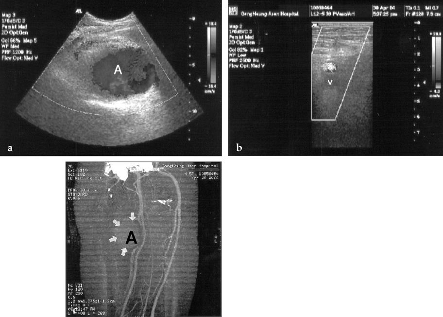
Fig. 1 (a) Preoperative color Doppler ultrasound showed a huge deep femoral artery aneurysm (A), measuring 8.5- × 9.0- × 7.5-cm. (b) The deep femoral artery aneurysm compressed the superficial femoral vein (v) resulting in venous stasis with suspicious soft thrombus formation in the right lower extremity veins (a; superficial femoral artery). (c) Preoperative multi-detector CT scan revealed a large pseudoaneurysm (A, black arrows) arising from the deep femoral artery. There was no evidence of aneurysms or occlusive lesions in the other arteries.
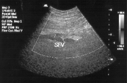
Fig. 2 Postoperative color Doppler ultrasound showed patent superficial femoral (SFV) and popliteal veins.
Reference
-
1. Cutler BS, Darling RC. Surgical management of arteriosclerotic femoral aneurysms. Surgery. 1973. 74:764–773.2. Roseman JM, Wyche D. True aneurysm of the profunda femoris artery. Literature review, differential diagnosis, management. J Cardiovasc Surg. 1987. 28:701–705.3. Johnson CA, Goff JM, Rehrig ST, Hadro NC. Asymptomatic profunda femoris artery aneurysm: diagnosis and rationale for management. Eur J Vasc Endovasc Surg. 2002. 24:91–92.4. Toda R, Yuda T, Watanabe S, Hisashi Y, Moriyama Y, Taira A. Surgical repair of a solitary deep femoral arterial aneurysm: report of two cases. Surg Today. 2000. 30:481–483.5. Valiulis AP, Johnston KW. Isolated atherosclerotic aneurysm of the profunda femoris artery. J Cardiovasc Surg (Torino). 1980. 21:498–500.6. Ratto GB, Sacco A, Canepa G, Motta G. Atherosclerotic aneurysm of the deep femoral artery. J Cardiovasc Surg (Torino). 1984. 25:574–576.7. Levi N, Schroeder TV. Arteriosclerotic femoral artery aneurysms. A short review. J Cardiovasc Surg (Torino). 1997. 38:335–338.8. Tait WF, Vohra RK, Carr HM, Thomson GJ, Walker MG. True profunda femoris aneurysms: are they more dangerous than other atherosclerotic aneurysms of the femoropopliteal segment? Ann Vasc Surg. 1991. 5:92–95.9. Burchi C, Cavallaro G, Amato D, Cavallaro A. Isolated true atherosclerotic aneurysm of the profunda femoris artery. Case Report. J Cardiovasc Surg (Torino). 1999. 40:577–581.10. Catalano C, Fraioli F, Laghi A, Napoli A, Bezzi M, Pediconi F, et al. Infrarenal aortic and lower-extremity arterial disease: diagnostic performance of multi-detector row CT angiography. Radiology. 2004. 231:555–563.
- Full Text Links
-
- Actions
-
Cited
- CITED
-
- Close
- Share
-
- Similar articles
-
- An Isolated Femoral Artery Aneurysm Presenting with Leg Swelling
- The True Deep Femoral Artery Aneurysm: A Case Report
- Rupture of Giant Superficial Femoral Artery Aneurysm in a Leukemic Patient Submitted to Chemotherapy
- Mycotic Aneurysm of the Left Subclavian Artery Presenting as Mediastinal Abscess: Case Report
- Acute Embolic Attack on Persistent Sciatic Artery: 1 case
