An Unusual Odontogenic Cyst with Diverse Histologic Features
- Affiliations
-
- 1Department of Oral Pathology, BK 21 Project, School of Dentistry, Chosun University, Gwangju, Korea. jhyoon@chosun.ac.kr
- 2Department of Oral and Maxillofacial Surgery, BK 21 Project, School of Dentistry, Chosun University, Gwangju, Korea.
- 3Department of Oral Pathology, Oral Cancer Research Institute, Yonsei University College of Dentistry, Seoul, Korea.
- KMID: 1715881
- DOI: http://doi.org/10.3349/ymj.2006.47.1.122
Abstract
- An unusual odontogenic cyst, which was originally believed to be a clinical dentigerous cyst associated with an impacted mandibular third molar, was found histologically to demonstrate the characteristics of a glandular odontogenic cyst with para- and orthokeratinization. These histologic diversities were interpreted as a reflection of the pluripotentiality of the epithelial remnants of the mandibular third molars or dentigerous cyst epithelium. It is possible that it has the capacity to induce the formation of cysts in both squamous and glandular epithelium.
MeSH Terms
Figure
-
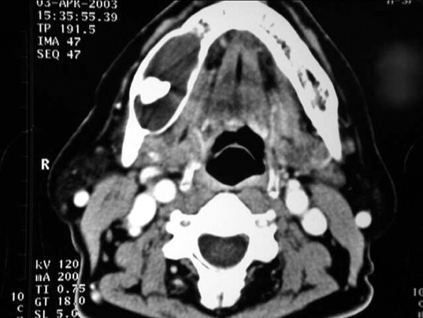
Fig. 1 Computed tomogram demonstrating a well demarcated cystic lesion with buccolingual expansion of the right mandible. A third molar tooth is impacted within the lesion.
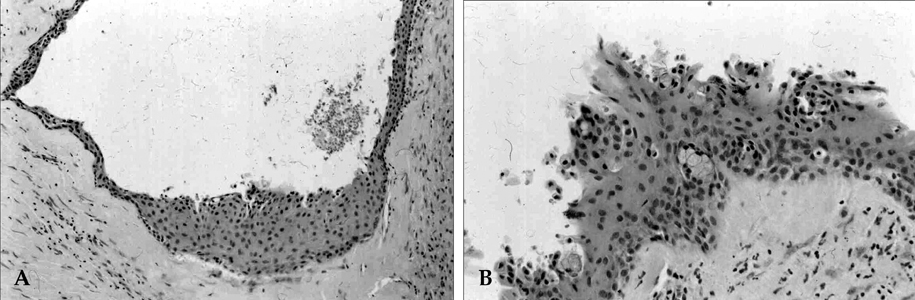
Fig. 2 A. Photomicrograph showing a non-keratinized squamous epithelium with cuboidal surface cells and an epithelial plaque of swirl-like arrangement (H-E, ×100). B. The cyst lining showing papillary projections, duct-like structures and clusters of goblet cells, resembling a glandular acinus (H-E, ×200).

Fig. 3 A. The lining epithelium is composed of a uniform layer of stratified squamous epithelium, with a hyperchromatic and palisaded basal cell layer. There is a corrugated parakeratotic surface, showing typical features of odontogenic keratocysts (H-E, ×200). B. The epithelial lining showing prominent keratohyaline granules beneath the orthokeratotic surface (H-E, ×200).
Reference
-
1. Pieuch JF, Eisenberg E, Segal D, Carlson R. Respiratory epithelium as an integral part of an odontogenic keratocyst: report of case. J Oral Surg. 1980. 38:445–447.2. Shear M. Cysts of the Oral Regions. 1992. 3rd ed. Oxford: Wright;4–50. 75–98. 136–178.3. High AS, Main DM, Khoo SP, Pedlar J, Hume WJ. The polymorphous odontogenic cyst. J Oral Pathol Med. 1996. 25:25–31.4. Gardner DG, Kessler HP, Morency R, Schaffner DL. The glandular odontogenic cyst: an apparent entity. J Oral Pathol. 1988. 17:359–366.5. Patron M, Colmenero C, Larrauri J. Glandular odontogenic cyst: clinicopathologic analysis of three cases. Oral Surg Oral Med Oral Pathol. 1991. 72:71–74.6. Marsland EA, Browne RM. Two odontogenic cysts, partially lined with ciliated epithelium. Oral Surg Oral Med Oral Pathol. 1965. 19:502–507.7. Browne RM. The odontogenic keratocyst. Histological features and their correlation with clinical behavior. Br Dent J. 1971. 131:249–259.8. Yamazaki M, Cheng J, Nomura T, Saito C, Hayashi T, Saku T. Maxillary odontogenic keratocyst with respiratory epithelium: a case report. J Oral Pathol Med. 2003. 32:496–498.9. Cutright DE. Histopathologic findings in third molar opercula. Oral Surg Oral Med Oral Pathol. 1976. 41:215–224.10. Browne RM. Metaplasia and degeneration in odontogenic cysts in man. J Oral Pathol. 1972. 1:145–158.11. Gorlin RJ. Potentialities of oral epithelium manifested by mandibular dentigerous cysts. Oral Surg Oral Med Oral Pathol. 1957. 10:271–284.
- Full Text Links
-
- Actions
-
Cited
- CITED
-
- Close
- Share
-
- Similar articles
-
- Calcifying Odontogenic Cyst Associated with an Impacted Upper Cuspid
- A Clinicostatistical Study Of Non-Odontogenic Cysts: The Experience Over 10 Years
- Recurrent Adenomatoid Odontogenic Tumor Arising From A Dentigerous Cyst
- Squamous cell carcinoma arising from residual odontogenic cyst: Report of a Case & Review of Literatures
- Glandular odontogenic cyst : Report of three cases
