J Korean Med Sci.
2007 Feb;22(1):156-158. 10.3346/jkms.2007.22.1.156.
Prenatal Detection of a Congenital Pancreatic Cyst by Ultrasound
- Affiliations
-
- 1Department of Obstetrics and Gynecology, College of Medicine, Chosun University, Gwangju, Korea. sjchoi@chosun.ac.kr
- 2Department of Pathology, College of Medicine, Chosun University, Gwangju, Korea.
- 3Department of Pediatric Surgery, College of Medicine, Chosun University, Gwangju, Korea.
- KMID: 1713258
- DOI: http://doi.org/10.3346/jkms.2007.22.1.156
Abstract
- We present a case of a fetal pancreatic cyst, a rare disease in fetal life, detected prenatally at 30 weeks' gestation by ultrasound. Routine ultrasound examination at 30 weeks' gestation by primary obstetrician showed a cyst on the fetal abdomen. Initially, the suspected diagnosis was a mesenteric cyst. Subsequent ultrasound examination at weeks 32, 36 showed a fetal retroperitoneal cyst. A 3.6 kg female neonate was born to 23 yr old woman by spontaneous vaginal delivery at 38 weeks' gestation. The fetus underwent exploratory laparotomy. Histopathologic and immunohistochemical diagnosis revealed the cyst to be a pancreatic cyst. Surgical outcome was excellent. Thus, we report this case of a pancreatic cyst detected via prenatal ultrasonography.
MeSH Terms
Figure
-

Fig. 1 (A) Ultrasonographic image showing a unilocular cyst in the fetal abdomen at 30 weeks' gestation. (B) Magnetic resonance image (T-2-weighted sequence) showing a huge cystic mass with high signal density in the neonatal abdomen.

Fig. 2 (A) In operative finding, the cystic mass was in the retroperitoneum between the transverse colon and the pancreatic tail. (B) The cystic mass which was removed from distal pancreas.
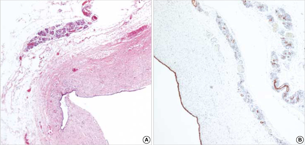
Fig. 3 (A) Histopathologic finding of the cyst showing the pancreatic tissue and lining epitheliums (H&E stain, ×40). (B) In immunohistochemical stain, cytokeratin is positive in the cystic epithelium and pancreatic tissue (×40).
Reference
-
1. Baker LL, Hartman GE, Northway WH. Sonographic detection of congenital pancreatic cysts in the newborn: report of a case and review of the literature. Pediatr Radiol. 1990. 20:488–490.
Article2. Auringer ST, Ulmer JL, Summer TE, Turner CS. Congenital cyst of the pancreas. J Pediatr Surg. 1993. 28:1570–1571.
Article3. Fremond B, Poulain P, Odent S, Milon J, Treguier C, Babut JM. Prenatal detection of a congenital pancreatic cyst and Beckwith-Wiedemann syndrome. Prenat Diagn. 1997. 17:276–280.4. Vane WD, Ashcraft WK, Holder MT. Lesions of the pancreas. Pediatric surgery. 1993. Philadelphia: Saunders;525–534.5. Dahnert W. Radiology review manual. 1996. Baltimore: Williams & Wilkins;591–593.6. Kebapci M, Aslan O, Kaya T, Ilhan H. Prenatal diagnosis of giant congenital pancreatic cyst of a neonate. Am J Roentgenol. 2000. 175:1408–1410.
Article7. Daher P, Diab N, Melki I, Abi-Aad G, Korkmaz G. Congenital cyst of the pancreas : antenatal diagnosis. Eur J Pediatr Surg. 1996. 6:180–182.
- Full Text Links
-
- Actions
-
Cited
- CITED
-
- Close
- Share
-
- Similar articles
-
- Prenatal Diagnosis and Outcome of Congenital Lung Mass
- Multiple Congenital Pancreatic Cysts in a Neonate
- Congenital lobar emphysema with cystic lung abnormality: Antenatal ultrasound appearance
- Prenatal Ultrasonographic Diagnosis of Frequent Congenital Fetal Anomalies
- Prenatal detection of fetal gastric duplication cyst: A case report
