Korean J Radiol.
2014 Feb;15(1):173-177. 10.3348/kjr.2014.15.1.173.
Spontaneous Intramural Full-Length Dissection of Esophagus Treated with Surgical Intervention: Multidetector CT Diagnosis with Multiplanar Reformations and Virtual Endoscopic Display
- Affiliations
-
- 1Department of Radiology, Soonchunhyang University Hospital Bucheon, Bucheon 420-767, Korea. acarad@naver.com
- 2Department of Thoracic and Cardiovascular Surgery, Soonchunhyang University Hospital Bucheon, Bucheon 420-767, Korea.
- KMID: 1711495
- DOI: http://doi.org/10.3348/kjr.2014.15.1.173
Abstract
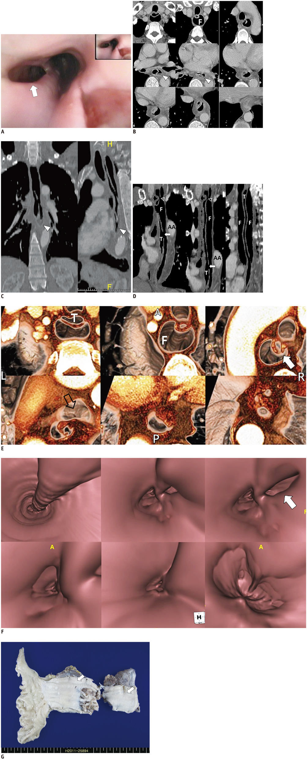
- Intramural esophageal dissection (IED) is an uncommon disorder characterized by separation of the mucosal and submucosal layers of the esophagus. Iatrogenic intervention is the most common cause of IED, but spontaneous dissection is rare. We report an unusually complicated case of spontaneous IED that involved the full-length of the esophagus that necessitated surgical intervention due to infection of the false lumen. In this case, chest computed tomography successfully established the diagnosis and aided in pre-operative evaluation with the use of various image post-processing techniques.
MeSH Terms
Figure
Reference
-
1. Krishnama MS, Ramadan MF, Curtisa J. Intramural esophageal dissection: CT imaging features. Eur J Radiol (extra). 2005; 56:17–19.2. Chiu HH, Lee SY. Intramural dissection of the esophagus endoscopic findings. J Intern Med Taiwan. 2006; 17:302–305.3. Marks IN, Keet AD. Intramural rupture of the oesophagus. Br Med J. 1968; 3:536–537.4. Kim SH, Lee SO. Circumferential intramural esophageal dissection successfully treated by endoscopic procedure and metal stent insertion. J Gastroenterol. 2005; 40:1065–1069.5. Soulellis CA, Hilzenrat N, Levental M. Intramucosal esophageal dissection leading to esophageal perforation: case report and review of the literature. Gastroenterol Hepatol (N Y). 2008; 4:362–365.6. Young CA, Menias CO, Bhalla S, Prasad SR. CT features of esophageal emergencies. Radiographics. 2008; 28:1541–1553.7. Hsu CC, Changchien CS. Endoscopic and radiological features of intramural esophageal dissection. Endoscopy. 2001; 33:379–381.8. Wu HC, Hsia JY, Hsu CP. Esophageal laceration with intramural dissection mimics esophageal perforation. Interact Cardiovasc Thorac Surg. 2008; 7:864–865.9. Kim SH, Lee JM, Han JK, Kim YH, Lee JY, Lee HJ, et al. Three-dimensional MDCT imaging and CT esophagography for evaluation of esophageal tumors: preliminary study. Eur Radiol. 2006; 16:2418–2426.10. Fadoo F, Ruiz DE, Dawn SK, Webb WR, Gotway MB. Helical CT esophagography for the evaluation of suspected esophageal perforation or rupture. AJR Am J Roentgenol. 2004; 182:1177–1179.
- Full Text Links
- Actions
-
Cited
- CITED
-
- Close
- Share
- Similar articles
-
- Spontaneous Intramural Hematoma of the Esophagus
- Endoscopic Treatment of Spontaneous Intramural Dissection of the Esophagus: A Case Report
- A Case of Spontaneous Intramural Hematoma of the Esophagus
- Primary Repair of Spontaneous Intramural Esophageal Dissection: A Case Report
- Intramural Esophageal Abscess and Dissection due to Retropharygeal Abscess


