Imaging Findings Of Desmoplastic Fibroma Rarely Involving The Clavicle: Case Report
- Affiliations
-
- 1Department of Radiology, Wonkwang University School of Medicine & Hospital, Iksan 570-749, Korea. juhngsk@wonkwang.ac.kr
- 2Department of Pathology, Wonkwang University School of Medicine & Hospital, Iksan 570-749, Korea.
- KMID: 1711487
- DOI: http://doi.org/10.3348/kjr.2014.15.1.130
Abstract
- Desmoplastic fibroma of bone is a rare locally aggressive, but non-metastatic tumor. In this case report, we present a desmoplastic fibroma in an unusual location, the clavicle. Desmoplastic fibroma involving the clavicle is extremely rare, with only 2 reported cases before 1985. We report the imaging findings of a desmoplastic fibroma of the clavicle with a review of the relevant literature.
Keyword
MeSH Terms
Figure
-
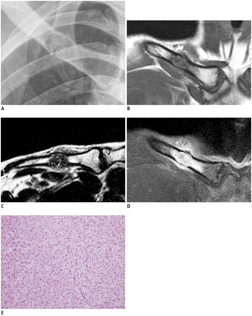
Fig. 1 Desmoplastic fibroma of clavicle in 28-year-old man. A. Part of plain anteroposterior radiograph of right clavicle reveals well-defined non-sclerotic marginated geographic lesion on medial one-third of right clavicle, with pathologic fracture. B-D. Coronal (B) T1-weighted magnetic resonance image shows hypointense to isointense signal compared to adjacent muscle, and lesion also demonstrates hypointense to isointense signal on axial (C) T2-weighted image. Gadolinium enhanced T1-weighted coronal (D) image reveals heterogeneous enhancement of mass. Also, adjacent bone marrow and soft tissue along periosteum are enhanced. E. Microscopic examination shows bland fibroblasts in background of numerous thick and wavy collagen fibers without evidence of cellular pleomorhpism, changes in nuclear-cytoplasmic ratio or mitotic activity (H&E, × 200).
Reference
-
1. Jaffe HL. Tumors and tumorous conditions of the bones and joints. Philadelphia, PA: Lea and Febiger;1958. p. 298–303.2. Gebhardt MC, Campbell CJ, Schiller AL, Mankin HJ. Desmoplastic fibroma of bone. A report of eight cases and review of the literature. J Bone Joint Surg Am. 1985; 67:732–747.3. Böhm P, Kröber S, Greschniok A, Laniado M, Kaiserling E. Desmoplastic fibroma of the bone. A report of two patients, review of the literature, and therapeutic implications. Cancer. 1996; 78:1011–1023.4. Smith J, Yuppa F, Watson RC. Primary tumors and tumor-like lesions of the clavicle. Skeletal Radiol. 1988; 17:235–246.5. Pratt GF, Dahlin DC, Ghormley RK. Tumors of the scapula and clavicle. Surg Gynecol Obstet. 1958; 106:536–544.6. Inwards CY, Unni KK, Beabout JW, Sim FH. Desmoplastic fibroma of bone. Cancer. 1991; 68:1978–1983.7. Crim JR, Gold RH, Mirra JM, Eckardt JJ, Bassett LW. Desmoplastic fibroma of bone: radiographic analysis. Radiology. 1989; 172:827–832.8. Taconis WK, Schütte HE, van der Heul RO. Desmoplastic fibroma of bone: a report of 18 cases. Skeletal Radiol. 1994; 23:283–288.9. Frick MA, Sundaram M, Unni KK, Inwards CY, Fabbri N, Trentani F, et al. Imaging findings in desmoplastic fibroma of bone: distinctive T2 characteristics. AJR Am J Roentgenol. 2005; 184:1762–1767.10. Vanhoenacker FM, Hauben E, De Beuckeleer LH, Willemen D, Van Marck E, De Schepper AM. Desmoplastic fibroma of bone: MRI features. Skeletal Radiol. 2000; 29:171–175.
