A Case of an Idiopathic Massive Osteolysis with Skip Lesions
- Affiliations
-
- 1Department of Radiology, Arnavutkoy State Hospital, Instanbul 34275, Turkey. drmozbayrak@hotmail.com
- 2Department of Radiology, Cerrahpasa Medical Faculty, Istanbul University, Istanbul 34452, Turkey.
- 3Department of Orthopedic Surgery, Cerrahpasa Medical Faculty, Istanbul University, Istanbul 34452, Turkey.
- 4Department of Pathology, Cerrahpasa Medical Faculty, Istanbul University, Istanbul 34452, Turkey.
- KMID: 1711462
- DOI: http://doi.org/10.3348/kjr.2013.14.6.946
Abstract
- A patient with a 2-year history of pain in the left arm, and decreased strengths unrelieved by non-steroidal anti-inflammatory therapy, was being referred for repeating radiography. The radiologic examinations have demonstrated a unique pattern of non-contiguous osteolysis in the left elbow, proximal and distal radius, ulna, wrist, carpal bones, proximal and distal metacarpals and phalanges. Multi-site biopsies were being performed and confirmed the diagnosis of massive osteolysis. To our knowledge, this is the first case in which multifocal, non-contiguous osteolysis with skip lesions without associated nephropathy and without a hereditary pattern is being described in one extremity.
MeSH Terms
Figure
-
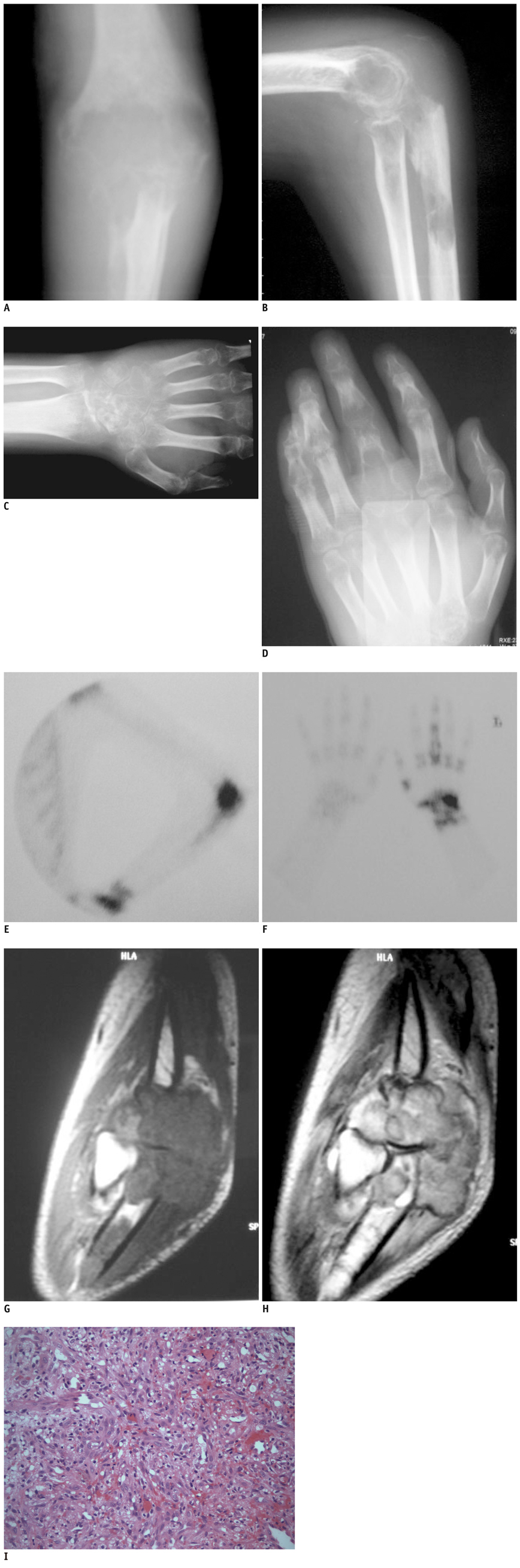
Fig. 1 Massive osteolysis with skip lesions in 38-year-old man. Plain radiography of upper left extremity demonstrate almost complete osteolysis of left elbow (A), proximal radius (B), distal radius and ulna, wrist and carpal bones (C), proximal and distal metacarpals and phalanges (D). Bone scintigraphy showed increased activities in osteolysed region: elbow (E), wrist and carpal bones and phalanges (F). Sagittal MR images demonstrated that soft tissue masses and affected bones were hypointense to isointense with muscle on T1-weighted imaging (G) and were of heterogenous signal intensity on T2-weighted imaging (H). (I) Photomicrograph (Hematoxylin and eosin, original magnification × 200) revealed fibrous tissue containing proliferated vascular structures replaced with bone trabeculae and bone marrow.
Reference
-
1. Jackson JBS. A boneless arm. Boston Med Surg J. 1838; 18:368–369.2. Jackson JBS. Absorption of the humerus after fracture. Boston Med Surg J. 1872; 10:245–247.3. Gorham LW, Stout AP. Massive osteolysis (acute spontaneous absorption of bone, phantom bone, disappearing bone); its relation to hemangiomatosis. J Bone Joint Surg Am. 1955; 37-A:985–1004.4. Hardegger F, Simpson LA, Segmueller G. The syndrome of idiopathic osteolysis. Classification, review, and case report. J Bone Joint Surg Br. 1985; 67:88–93.5. Yoo SY, Hong SH, Chung HW, Choi JA, Kim CJ, Kang HS. MRI of Gorham's disease: findings in two cases. Skeletal Radiol. 2002; 31:301–306.6. Sato K, Sugiura H, Yamamura S, Mieno T, Nagasaka T, Nakashima N. Gorham massive osteolysis. Arch Orthop Trauma Surg. 1997; 116:510–513.7. Resnick D, Kransdorf MJ. Bone and joint imaging. 3rd ed. Philadelphia: Elsevier Saunders;2005. p. 1449–1451.8. White AA 3rd. Disappearing bone disease with arthropathy and severe scarring of the skin. A report of four cases seen in South Vietnam. J Bone Joint Surg Br. 1971; 53:303–309.9. Beals RK, Bird CB. Carpal and tarsal osteolysis. A case report and review of the literature. J Bone Joint Surg Am. 1975; 57:681–686.10. Tookman AG, Paice EW, White AG. Idiopathic multicentric osteolysis with acro-osteolysis. A case report. J Bone Joint Surg Br. 1985; 67:86–88.11. Tauro B. Multicentric Gorham's disease. J Bone Joint Surg Br. 1992; 74:928–929.12. Downing ND, Garnavos C, Lunn PG. Idiopathic multicentric osteolysis principally affecting the phalanges of the hands and feet. J Hand Surg Br. 1996; 21:656–659.13. Burkhard O, Beyer J, Schrezenmeir J, Brunier A, Delling G. [A case of idiopathic multicentric osteolysis]. Med Klin (Munich). 1989; 84:364–368.14. Johnson PM, McClure JG. Observations on massive osteolysis; a review of the literature and report of a case. Radiology. 1958; 71:28–42.
- Full Text Links
-
- Actions
-
Cited
- CITED
-
- Close
- Share
-
- Similar articles
-
- Band acro-osteolysis in a Black woman: a case report and review of the literature
- Gorham's Syndrom: A Case Report
- Diffuse Neonatal Hemangiomatosis with Association of Massive Osteolysis and Arteriovenous Fistulae: An autopsy case
- Diffuse Neonatal Hemangiomatosis with Association of Massive Osteolysis and Arteriovenous Fistulae: An autopsy case
- Treatment of Pelvic Osteolysis with a stable Cementless Acetabular Cup with Exchange of Polyethlene Liner and Morselized Bone Graft
