MR Imaging Findings of Extraovarian Endocervical Mucinous Borderline Tumors Arising from Pelvic Endometriosis
- Affiliations
-
- 1Department of Radiology, Seoul St. Mary's Hospital, The Catholic University of Korea, Seoul 137-701, Korea. serha@catholic.ac.kr
- 2Department of Hospital Pathology, Seoul St. Mary's Hospital, The Catholic University of Korea, Seoul 137-701, Korea.
- 3Department of Obstetrics and Gynecology, Seoul St. Mary's Hospital, The Catholic University of Korea, Seoul 137-701, Korea.
- KMID: 1711458
- DOI: http://doi.org/10.3348/kjr.2013.14.6.918
Abstract
- We report MR imaging findings of a rare case of endocervical mucinous borderline tumor (MBT) involving the cul-de-sac and left fallopian tube arising from extensive pelvic endometriosis with pathologic correlation in a 35-year-old woman presented with vague pelvic pain. Endocervical MBT is a type of endometriosis-associated carcinoma. Imaging findings of endocervical MBT are unilocular or oligolocular cystic lesions with enhancing mural nodules, which are different from those of the more common intestinal type MBT.
MeSH Terms
Figure
-
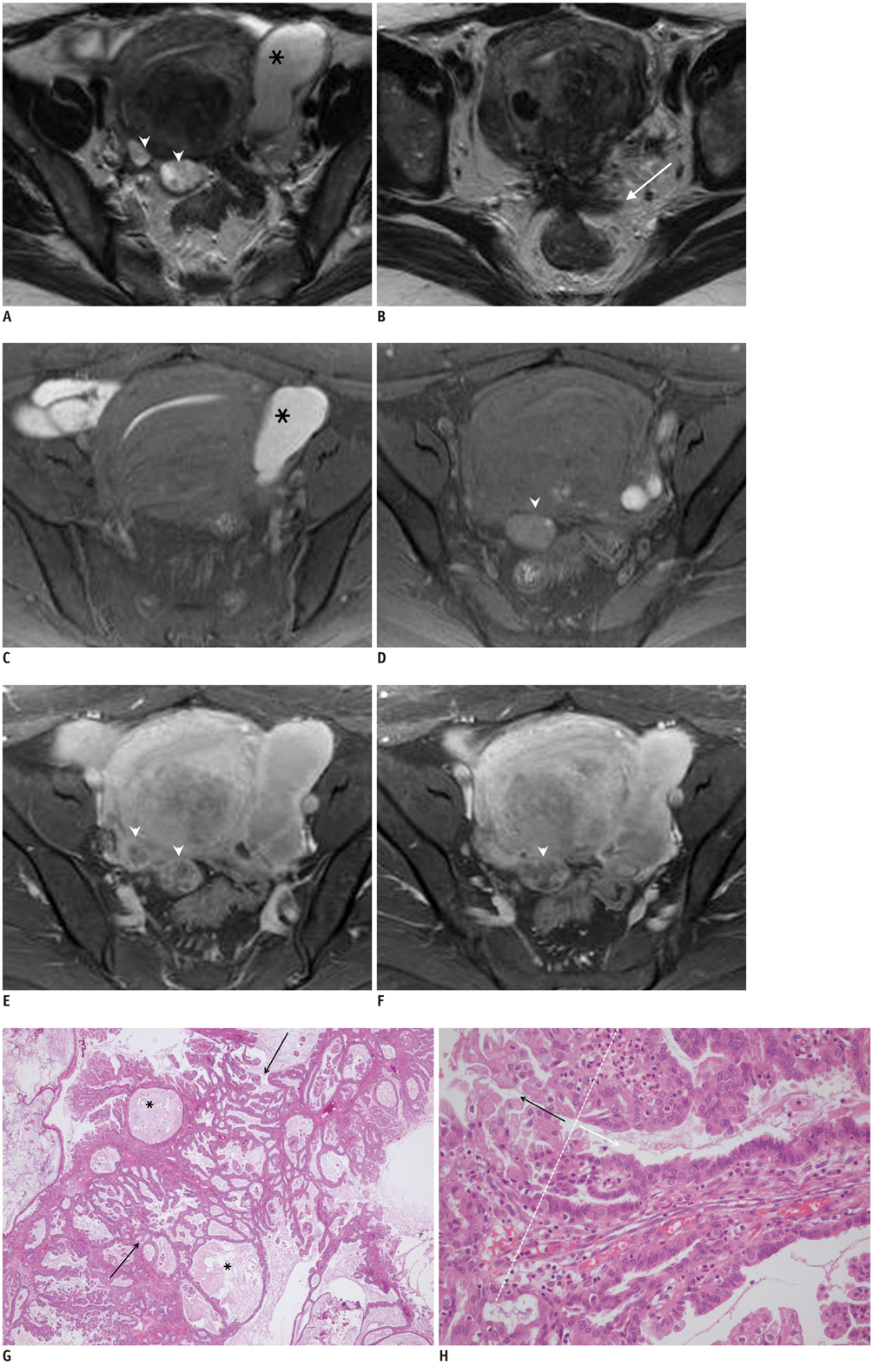
Fig. 1 Thirty five-year-old woman with extraovarian endocervical mucinous borderline tumor arising from pelvic endometriosis. Consecutive axial T2-weighted (A, B), fat-suppressed T1-weighted (C, D) and contrast-enhanced fat-suppressed T1-weighted (E, F) MR images. MR images show elongated tubular cystic lesion (asterisk in A, C) with high signal intensity on both T1- and T2-weighted images in left adnexa, which is diagnosed as hematosalpinx. Axial T2-weighted image (B) shows stellate low signal intensity lesion (arrow in B) with obliteration of cul-de-sac, suggestive of deep infiltrating endometriosis. Uterus is diffusely enlarged with focal adenomyosis and intramural myoma in posterior wall. Two small cystic lesions (arrowheads in A, D) with high signal intensity on T2-weighted image and slightly high signal intensity on T1-weighted image are present in cul-de-sac along posterior margin of uterus. Following contrast-infusion, peripheral nodular enhancement is noted in cystic lesions (arrowheads in E, F), raising possibility of malignancy. Histopathologic findings of cul-de-sac masses show multiple mucin-filled cysts (asterisks in G) and papillary projections (arrows in G) lined by endocervical type mucinous cells. These masses were confirmed as endocervical mucinous borderline tumors. Gradual transition from epitheliums of endometriosis (white arrow in H) to mucinous tumor cells (black arrow in H) was identified.
Reference
-
1. Melin A, Sparén P, Persson I, Bergqvist A. Endometriosis and the risk of cancer with special emphasis on ovarian cancer. Hum Reprod. 2006; 21:1237–1242.2. Stern RC, Dash R, Bentley RC, Snyder MJ, Haney AF, Robboy SJ. Malignancy in endometriosis: frequency and comparison of ovarian and extraovarian types. Int J Gynecol Pathol. 2001; 20:133–139.3. Yoshikawa H, Jimbo H, Okada S, Matsumoto K, Onda T, Yasugi T, et al. Prevalence of endometriosis in ovarian cancer. Gynecol Obstet Invest. 2000; 50:Suppl 1. 11–17.4. Kataoka M, Togashi K, Koyama T, Yamaoka T, Iwasa Y, Fujii S, et al. MR imaging of müllerian mucinous borderline tumors arising from endometriotic cysts. J Comput Assist Tomogr. 2002; 26:532–537.5. McDermott S, Oei TN, Iyer VR, Lee SI. MR imaging of malignancies arising in endometriomas and extraovarian endometriosis. Radiographics. 2012; 32:845–863.6. Bedaiwy MA, Hussein MR, Biscotti C, Falcone T. Pelvic endometriosis is rarely associated with ovarian borderline tumours, cytologic and architectural atypia: a clinicopathologic study. Pathol Oncol Res. 2009; 15:81–88.7. Brooks JJ, Wheeler JE. Malignancy arising in extragonadal endometriosis: a case report and summary of the world literature. Cancer. 1977; 40:3065–3073.8. Benoit L, Arnould L, Cheynel N, Diane B, Causeret S, Machado A, et al. Malignant extraovarian endometriosis: a review. Eur J Surg Oncol. 2006; 32:6–11.9. Scott RB. Malignant changes in endometriosis. Obstet Gynecol. 1953; 2:283–289.10. Kim KR, Choi J, Hwang JE, Baik YA, Shim JY, Kim YM, et al. Endocervical-like (Müllerian) mucinous borderline tumours of the ovary are frequently associated with the KRAS mutation. Histopathology. 2010; 57:587–596.11. Shappell HW, Riopel MA, Smith Sehdev AE, Ronnett BM, Kurman RJ. Diagnostic criteria and behavior of ovarian seromucinous (endocervical-type mucinous and mixed cell-type) tumors: atypical proliferative (borderline) tumors, intraepithelial, microinvasive, and invasive carcinomas. Am J Surg Pathol. 2002; 26:1529–1541.12. Rutgers JL, Scully RE. Ovarian mullerian mucinous papillary cystadenomas of borderline malignancy. A clinicopathologic analysis. Cancer. 1988; 61:340–348.13. Tanaka YO, Okada S, Yagi T, Satoh T, Oki A, Tsunoda H, et al. MRI of endometriotic cysts in association with ovarian carcinoma. AJR Am J Roentgenol. 2010; 194:355–361.
- Full Text Links
-
- Actions
-
Cited
- CITED
-
- Close
- Share
-
- Similar articles
-
- MR Findings of Extrauterine Mullerian Adenosarcoma Associated with Deep Pelvic Endometriosis
- A Case of Ovarian Mullerian Mucinous Papillary Cystadenoma of Borderline Malignancy
- Ovarian Large Cell Neuroendocrine Carcinoma Associated with Endocervical-like Mucinous Borderline Tumor: A Case Report and Literature Review
- Morphometric Study on Mucinous Tumors of the Ovary
- A Case of Ovarian Mullerian Mucinous Papillary Cystadenoma of Borderline Malignancy
