Biliary Peritonitis after Radiofrequency Ablation Diagnosed by Gadoxetic Acid-Enhanced MR Imaging
- Affiliations
-
- 1Department of Diagnostic Imaging and Nuclear Medicine, Kyoto University Graduate School of Medicine, Kyoto 606-8507, Japan. akihirof@kuhp.kyoto-u.ac.jp
- 2Department of Radiology, Osaka Red Cross Hospital, Osaka 543-8555, Japan.
- 3Department of Gastroenterology and Hepatology, Osaka Red Cross Hospital, Osaka 543-8555, Japan.
- KMID: 1711457
- DOI: http://doi.org/10.3348/kjr.2013.14.6.914
Abstract
- This study describes the first case of biliary peritonitis after radiofrequency ablation diagnosed by magnetic resonance (MR) imaging with gadolinium ethoxybenzyl diethylenetriaminepentaacetic acid (Gd-EOB-DTPA), a hepatocyte-specific MR imaging contrast agent. The image acquired 300 minutes after the administration of Gd-EOB-DTPA was useful to make a definite diagnosis and to identify the pathway of bile leakage. It is important to decide on the acquisition timing with consideration of the predicted location of bile duct injury.
Keyword
MeSH Terms
-
Aged, 80 and over
Bile Duct Diseases/*diagnosis/etiology
Carcinoma, Hepatocellular/diagnosis/surgery
Catheter Ablation/*adverse effects
Contrast Media/diagnostic use
Diagnosis, Differential
Follow-Up Studies
Gadolinium DTPA/*diagnostic use
Hepatectomy/adverse effects/methods
Humans
Liver Neoplasms/diagnosis/*surgery
Magnetic Resonance Imaging/*methods
Male
Peritonitis/*diagnosis/etiology
Contrast Media
Gadolinium DTPA
Figure
-
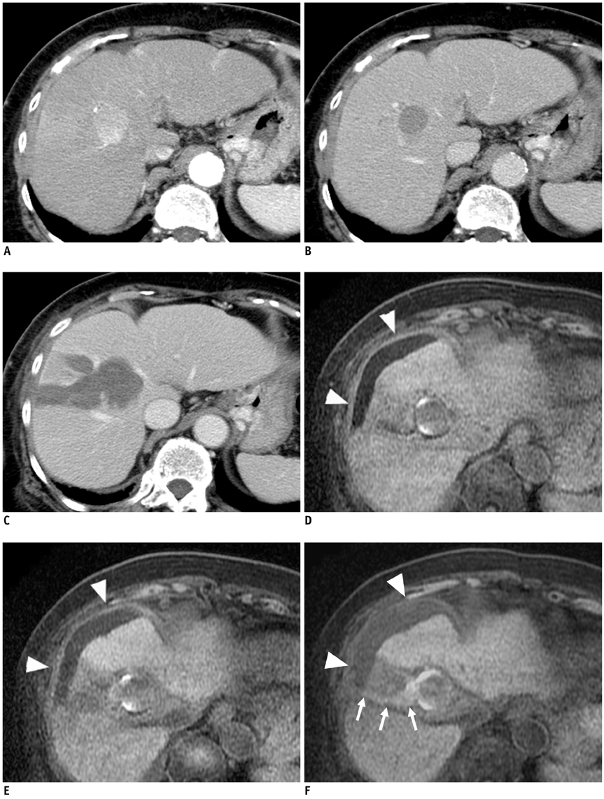
Fig. 1 Dynamic contrast-enhanced CT before RFA, contrast-enhanced CT after RFA, and Gd-EOB-DTPA-enhanced MRI two months after RFA in 80-year-old woman. Dynamic contrast-enhanced CT images show 3-cm hepatic mass in segment VIII, which has marked enhancement in arterial phase (A) and low attenuation compared to liver parenchyma in portal venous phase (B) before RFA. Contrast-enhanced CT obtained seven days after RFA reveals ablated tumor with sufficient margin (C). Two months after RFA, enhanced T1-weighted images were acquired at 20 minutes (D), 60 minutes (E), and 300 minutes (F) after administration of Gd-EOB-DTPA. Enhanced T1-weighted MR imaging acquired at 300 minutes shows increased signal intensity in fluid collection around liver than that acquired at 20 and 60 minutes (arrowheads). Linear high signal areas to match RFA puncture route are also demonstrated in enhanced T1-weighted MR imaging at 300 minutes (arrows). RFA = radiofrequency ablation, Gd-EOB-DTPA = adolinium ethoxybenzyl diethylenetriaminepentaacetic acid
Reference
-
1. Karabulut N, Cakmak V, Kiter G. Confident diagnosis of bronchobiliary fistula using contrast-enhanced magnetic resonance cholangiography. Korean J Radiol. 2010; 11:493–496.2. Lee NK, Kim S, Lee JW, Lee SH, Kang DH, Kim GH, et al. Biliary MR imaging with Gd-EOB-DTPA and its clinical applications. Radiographics. 2009; 29:1707–1724.3. Aduna M, Larena JA, Martín D, Martínez-Guereñu B, Aguirre I, Astigarraga E. Bile duct leaks after laparoscopic cholecystectomy: value of contrast-enhanced MRCP. Abdom Imaging. 2005; 30:480–487.4. Tateishi R, Shiina S, Teratani T, Obi S, Sato S, Koike Y, et al. Percutaneous radiofrequency ablation for hepatocellular carcinoma. An analysis of 1000 cases. Cancer. 2005; 103:1201–1209.5. Chang IS, Rhim H, Kim SH, Kim YS, Choi D, Park Y, et al. Biloma formation after radiofrequency ablation of hepatocellular carcinoma: incidence, imaging features, and clinical significance. AJR Am J Roentgenol. 2010; 195:1131–1136.6. Chen MH, Yang W, Yan K, Gao W, Dai Y, Wang YB, et al. Treatment efficacy of radiofrequency ablation of 338 patients with hepatic malignant tumor and the relevant complications. World J Gastroenterol. 2005; 11:6395–6401.7. Bollow M, Taupitz M, Hamm B, Staks T, Wolf KJ, Weinmann HJ. Gadolinium-ethoxybenzyl-DTPA as a hepatobiliary contrast agent for use in MR cholangiography: results of an in vivo phase-I clinical evaluation. Eur Radiol. 1997; 7:126–132.8. Tschirch FT, Struwe A, Petrowsky H, Kakales I, Marincek B, Weishaupt D. Contrast-enhanced MR cholangiography with Gd-EOB-DTPA in patients with liver cirrhosis: visualization of the biliary ducts in comparison with patients with normal liver parenchyma. Eur Radiol. 2008; 18:1577–1586.9. Pasmans HL, Go PM, Gouma DJ, Heidendal GA, van Engelshoven JM, van Kroonenburgh MJ. Scintigraphic diagnosis of bile leakage after laparoscopic cholecystectomy. A prospective study. Clin Nucl Med. 1992; 17:697–700.10. Dinkel HP, Moll R, Gassel HJ, Knüpffer J, Timmermann W, Fieger M, et al. Helical CT cholangiography for the detection and localization of bile duct leakage. AJR Am J Roentgenol. 1999; 173:613–617.
- Full Text Links
-
- Actions
-
Cited
- CITED
-
- Close
- Share
-
- Similar articles
-
- Evaluation and Prediction of Post-Hepatectomy Liver Failure Using Imaging Techniques: Value of Gadoxetic Acid-Enhanced Magnetic Resonance Imaging
- Current Limitations and Potential Breakthroughs for the Early Diagnosis of Hepatocellular Carcinoma
- Hepatic Angiomyolipoma Presenting as a Hyperintense Lesion During the Hepatobiliary Phase of Gadoxetic Acid Enhanced-MRI: a Case Report
- Diagnosis of Hepatocellular Carcinoma with Gadoxetic Acid-Enhanced MRI: 2016 Consensus Recommendations of the Korean Society of Abdominal Radiology
- Gadoxetic acid-enhanced magnetic resonance imaging: Hepatocellular carcinoma and mimickers
