Korean J Ophthalmol.
2013 Oct;27(5):392-395. 10.3341/kjo.2013.27.5.392.
Surgical Removal of Retained Subfoveal Perfluorocarbon Liquid through a Therapeutic Macular Hole with Intravitreal PFCL Injection and Gas Tamponade
- Affiliations
-
- 1Department of Ophthalmology, Seoul National University Hospital, Seoul National University College of Medicine, Seoul, Korea.
- 2Department of Ophthalmology, Seoul National University Bundang Hospital, Seoul National University College of Medicine, Seongnam, Korea. sejoon1@daum.net
- 3Seoul Artificial Eye Center, Seoul National University Hospital Clinical Research Institute, Seoul, Korea.
- KMID: 1707297
- DOI: http://doi.org/10.3341/kjo.2013.27.5.392
Abstract
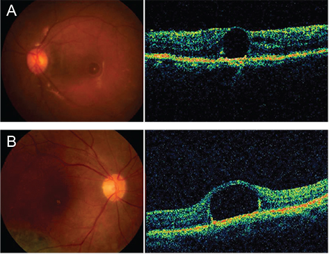
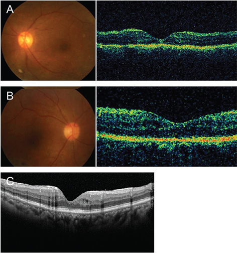
- We report two cases of surgical removal of a retained subfoveal perfluorocarbon liquid (PFCL) bubble through a therapeutic macular hole combined with intravitreal PFCL injection and gas tamponade. Two patients underwent pars plana vitrectomy with PFCL injection for rhegmatogenous retinal detachment. In both cases, a retained subfoveal PFCL bubble was noticed postoperatively by funduscopy and optical coherence tomography. Both patients underwent surgical removal of the subfoveal PFCL through a therapeutic macular hole and gas tamponade. The therapeutic macular holes were completely closed by gas tamponade and the procedure yielded a good visual outcome (best-corrected visual acuity of 20 / 40 in both cases). In one case, additional intravitreal PFCL injection onto the macula reduced the size of the therapeutic macular hole and preserved the retinal structures in the macula. Surgical removal of a retained subfoveal PFCL bubble through a therapeutic macular hole combined with intravitreal PFCL injection and gas tamponade provides an effective treatment option.
MeSH Terms
Figure
Reference
-
1. Bourke RD, Simpson RN, Cooling RJ, Sparrow JR. The stability of perfluoro-N-octane during vitreoretinal procedures. Arch Ophthalmol. 1996; 114:537–544.2. Garcia-Valenzuela E, Ito Y, Abrams GW. Risk factors for retention of subretinal perfluorocarbon liquid in vitreoretinal surgery. Retina. 2004; 24:746–752.3. Berglin L, Ren J, Algvere PV. Retinal detachment and degeneration in response to subretinal perfluorodecalin in rabbit eyes. Graefes Arch Clin Exp Ophthalmol. 1993; 231:233–237.4. Lee GA, Finnegan SJ, Bourke RD. Subretinal perfluorodecalin toxicity. Aust N Z J Ophthalmol. 1998; 26:57–60.5. Lai JC, Postel EA, McCuen BW 2nd. Recovery of visual function after removal of chronic subfoveal perfluorocarbon liquid. Retina. 2003; 23:868–870.6. Lesnoni G, Rossi T, Gelso A. Subfoveal liquid perfluorocarbon. Retina. 2004; 24:172–176.7. Le Tien V, Pierre-Kahn V, Azan F, et al. Displacement of retained subfoveal perfluorocarbon liquid after vitreoretinal surgery. Arch Ophthalmol. 2008; 126:98–101.8. Roth DB, Sears JE, Lewis H. Removal of retained subfoveal perfluoro-n-octane liquid. Am J Ophthalmol. 2004; 138:287–289.9. Garcia-Arumi J, Castillo P, Lopez M, et al. Removal of retained subretinal perfluorocarbon liquid. Br J Ophthalmol. 2008; 92:1693–1694.
- Full Text Links
- Actions
-
Cited
- CITED
-
- Close
- Share
- Similar articles
-
- Complications caused by perfluorocarbon liquid used in pars plana vitrectomy
- Autologous Retinal Flap Transplantation of a Refractory Giant Macular Hole with Retinal Detachment
- Clinical Evaluation for Patients with Retinal Detachment Caused by Macular Hole
- A Case Report of Closed Traumatic Macular Hole after Intravit real Gas Injection
- The 2 Cases of Surgical Treatment for Retinal Detachment with Macular Hole after Ocular Trauma


