Korean J Radiol.
2014 Apr;15(2):185-187. 10.3348/kjr.2014.15.2.185.
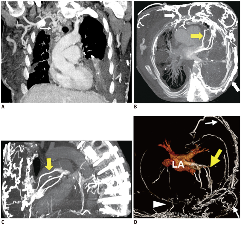
Superior Vena Cava Syndrome Associated with Right-to-Left Shunt through Systemic-to-Pulmonary Venous Collaterals
- Affiliations
-
- 1Department of Radiology, Brigham and Women's Hospital, Harvard Medical School, Boston, MA 02115, USA. yjuan@partners.org
- 2Department of Medical Imaging and Intervention, Chang Gung Memorial Hospital, Linkou and Chang Gung University, Taoyuan 333, Taiwan.
- 3Cardiology Division, Brigham and Women's Hospital, Harvard Medical School, Boston, MA 02115, USA.
- 4Department of Medical Imaging and Intervention, Chang Gung Memorial Hospital, Keelung and Chang Gung University, Keelung 204, Taiwan.
- KMID: 1705571
- DOI: http://doi.org/10.3348/kjr.2014.15.2.185
Abstract
- Superior vena cava (SVC) obstruction is associated with the gradual development of venous collaterals. We present a rare form of systemic-to-pulmonary subpleural collateral pathway that developed in the bridging subpleural pulmonary veins in a 54-year-old woman with complete SVC obstruction. This uncommon collateral pathway represents a rare form of acquired right-to-left shunt due to previous pleural adhesions with an increased risk of stroke due to right-to-left venous shunting, which requires lifelong anticoagulation.
MeSH Terms
Figure
Reference
-
1. Eren S, Karaman A, Okur A. The superior vena cava syndrome caused by malignant disease. Imaging with multi-detector row CT. Eur J Radiol. 2006; 59:93–103.2. Kim HC, Chung JW, Yoon CJ, Lee W, Jae HJ, Kim YI, et al. Collateral pathways in thoracic central venous obstruction: three-dimensional display using direct spiral computed tomography venography. J Comput Assist Tomogr. 2004; 28:24–33.3. Sze DY, Fleischmann D, Ma AO, Price EA, McConnell MV. Embolization of a symptomatic systemic to pulmonary (right-to-left) venous shunt caused by fibrosing mediastinitis and superior vena caval occlusion. J Vasc Interv Radiol. 2010; 21:140–143.4. Kapur S, Paik E, Rezaei A, Vu DN. Where there is blood, there is a way: unusual collateral vessels in superior and inferior vena cava obstruction. Radiographics. 2010; 30:67–78.5. Kim HC, Chung JW, Park SH, Kim HB, Jae HJ, Lee W, et al. Systemic-to-pulmonary venous shunt in superior vena cava obstruction: depiction on computed tomography venography. Acta Radiol. 2004; 45:269–274.6. Koetters KT. Superior vena cava syndrome. J Emerg Nurs. 2012; 38:135–138.7. Lawler LP, Fishman EK. Thoracic venous anatomy multidetector row CT evaluation. Radiol Clin North Am. 2003; 41:545–560.8. Sheth S, Ebert MD, Fishman EK. Superior vena cava obstruction evaluation with MDCT. AJR Am J Roentgenol. 2010; 194:W336–W346.9. Kim HJ, Kim HS, Chung SH. CT diagnosis of superior vena cava syndrome: importance of collateral vessels. AJR Am J Roentgenol. 1993; 161:539–542.
- Full Text Links
- Actions
-
Cited
- CITED
-
- Close
- Share
- Similar articles
-
- Right to Left Shunt as a Collateral Circulation in a Patient with Superior Vena Cava Syndrome: A Case Report
- Reopening of the Left Superior Vena Cava in Right Superior Vena Cava Obstruction
- CT Demonstration of the Extracardiac Anastomoses of the Coronary Veins in Superior Vena Cava or Left Brachiocephalic Vein Obstruction
- Reconstruction of the Superior Vena Cava with Extra-luminal Bypass Shunt
- A Case of Superior Vena Cava Syndrome


