A New Examination Method for Anatomical Variations of the Flexor Digitorum Superficialis in the Little Finger
- Affiliations
-
- 1Department of Hand Surgery, Hand Surgery Research Center, Affiliated Hospital of Nantong University, Nantong, China.
- 2Department of Orthopaedic Surgery, Asan Medical Center, Ulsan University College of Medicine, Seoul, Korea. jeonchoi@gmail.com
- 3Department of Orthopedic Surgery, Kyungpook National University Hospital, Daegu, Korea.
- KMID: 1705536
- DOI: http://doi.org/10.4055/cios.2013.5.2.138
Abstract
- BACKGROUND
Current examination methods to assess the anatomical variations of flexor digitorum superficialis (FDS) tendon in the little finger necessitate a strong external force applied by the examiner and cause false negatives. A new examination method was designed to detect the variations more accurately.
METHODS
We examined the little fingers of 220 adult hands (110 subjects) by 2 methods: the expanded examination method advocated by Tan et al., and a new examination method. Variations of the FDS in the little finger were examined by both methods and categorized separately as having independent FDS function, FDS connection to the tendons of the ring finger or of the multiple adjacent fingers, and functional substitution of the flexor digitorum profundus (FDP) with or without tendinous connection to the ring or multiple adjacent fingers. By our new method, we could further divide the FDS connection or FDP substitution with connection to the ring finger into 2 subtypes: loose and close connections. Data were reported as case numbers and percent. Date on symmetry were statistically analyzed by matched case-control studies.
RESULTS
Among 220 hands, 113 hands (51.4%) had independent FDS function by the new examination method, which was lower than the incidence (55.5%) detected with the existing expanded examination method. In the hands with connections between FDS tendons of the little and the ring fingers, 32 hands (14.5%) demonstrated loose and 37 (16.8%) close connections. Three hands (1.4%) had loose and 19 (8.6%) had close FDP substitution with tendinous connection to the ring finger. Among 110 hands without independent FDS function, variants of 42 hands (38.2%) were asymmetric. There was no statistical significance in symmetry of variations.
CONCLUSIONS
This new examination method offers other assessment variations of FDS tendon in the little finger. We recommend using this test to assess the variations and function of the FDS of the little finger.
MeSH Terms
Figure
-
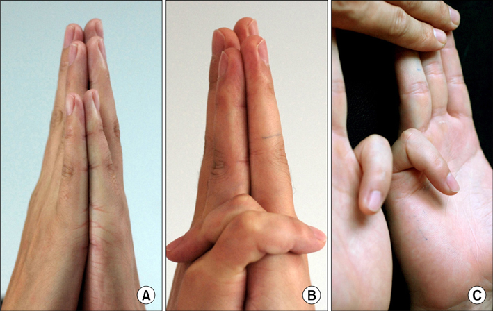
Fig. 1 Independent flexor digitorum superficialis function, examined by both the expanded and our new tests. (A) Bilateral palms and corresponding fingers are held facing each other in our new method. (B) The little fingers can touch the dorsal sides of both hands, with the proximal interphalangeal (PIP) joints flexed and the distal interphalangeal (DIP) joints extended in our new method. No gap between the ring fingers is seen. (C) The little fingers flex the PIP joint without DIP flexion by the expanded test.
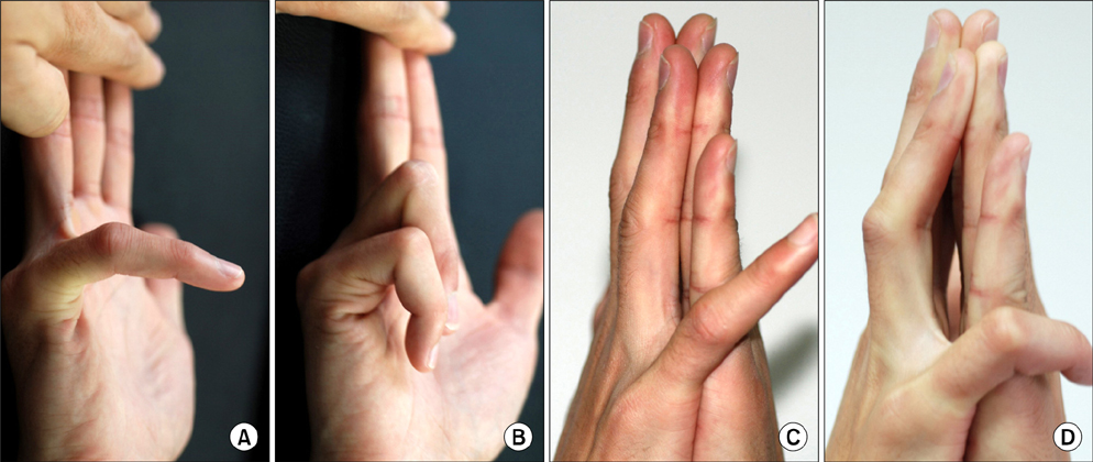
Fig. 2 Showing a connection of the flexor digitorum superficialis (FDS) between the little and ring fingers of the right hand by the expanded and our new tests. (A) Examination by the expanded test shows slight flexion at the proximal interphalangeal (PIP) joint, unless (B) releasing of the ring finger allows PIP joint of the little finger flexion. (C) Our new examination test shows when the two ring fingers keep straight and maintain closely touched, difficulty in flexing the PIP joint of the little finger to touch the target. (D) Mild flexion of the PIP joint of the adjacent ring finger was recorded as the loose FDS connection between the little and ring fingers in our new test.
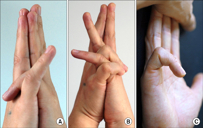
Fig. 3 The flexor digitorum superficialis (FDS) connection between the little and ring fingers of the right hand. (A) Inability of flexing the proximal interphalangeal (PIP) joint of the little finger of the right hand to touch the target by our new test, in contrast to the little finger of the left hand. (B) When the adjacent ring finger was permitted to flex together, the little finger could flex the PIP joint to reach the aim. Full flexion of the adjacent ring finger was recorded as the close connection between the little and ring fingers. (C) However, this case exhibited independent FDS function of the little finger when tested by the expanded method.

Fig. 4 The proximal interphalangeal (PIP) joint of the little finger could only be flexed in conjunction with the distal interphalangeal (DIP) joint flexion of the same finger, this was designated as flexor digitorum profundus (FDP) substitution (flexor digitorum superficialis [FDS] deficiency), including three types. 1) A&E showed the PIP joint of the little finger of the right hand flexed dependent on the FDP action of the same finger, but the FDP tendon of the little finger has no anatomical connection with the other fingers. 2) B&F (loose) and C&G (close) showed the FDS deficiency of the little finger of the right hand, and the FDP tendon of the little finger has connection with the FDS or FDP tendons of the adjacent ring finger. 3) D&H showed the FDS deficiency of the little finger of the left hand, and the FDP tendon of the little finger has connection with the flexor tendons of the ring and middle fingers. (A-D) were examined by the expanded method, and (E-H) by our new test.
Reference
-
1. Kaplan EB. Muscular and tendinous variations of the flexor superficialis of the fifth finger of the hand. Bull Hosp Joint Dis. 1969. 30(1):59–67.2. Shrewsbury MM, Kuczynski K. Flexor digitorum superficialis tendon in the fingers of the human hand. Hand. 1974. 6(2):121–133.
Article3. Gonzalez MH, Whittum J, Kogan M, Weinzweig N. Variations of the flexor digitorum superficialis tendon of the little finger. J Hand Surg Br. 1997. 22(2):277–280.
Article4. Christensen S. Anomalous muscle belly of the flexor digitorum superficialis in two generations. Hand. 1977. 9(2):162–164.
Article5. Elias LS, Schulter-Ellis FP. Anomalous flexor superficialis indicis: two case reports and literature review. J Hand Surg Am. 1985. 10(2):296–299.
Article6. Austin GJ, Leslie BM, Ruby LK. Variations of the flexor digitorum superficialis of the small finger. J Hand Surg Am. 1989. 14(2 Pt 1):262–267.
Article7. Baker DS, Gaul JS Jr, Williams VK, Graves M. The little finger superficialis: clinical investigation of its anatomic and functional shortcomings. J Hand Surg Am. 1981. 6(4):374–378.
Article8. Stein A, Lemos M, Stein S. Clinical evaluation of flexor tendon function in the small finger. Ann Emerg Med. 1990. 19(9):991–993.
Article9. Tan JS, Oh L, Louis DS. Variations of the flexor digitorum superficialis as determined by an expanded clinical examination. J Hand Surg Am. 2009. 34(5):900–906.
Article
- Full Text Links
-
- Actions
-
Cited
- CITED
-
- Close
- Share
-
- Similar articles
-
- Concomitant Variations in Flexor Digitorum Superficialis: A Case Report
- Carpal Tunnel Syndrome Caused by Anatomic Variation of Flexor Digitorum Superficialis of Little Finger
- A Case of Trigger Finger Following Longitudinal Tear of Flexor Digitorum Superficialis after Repeated Closed Injury
- "Trigger Finger at the Wrist" due to Anomalous Flexor Digitorum Superficialis Muscle Belly with Carpal Tunnel Syndrome: A Case Report
- Resection of one slip of the flexor digitorum superficialis resulting finger deformity in pediatric trigger finger: a case report
