Diffusion Weighted Imaging Findings in the Acute Lateral Medullary Infarction
- Affiliations
-
- 1Department of Neurology, Pundang Jesaeng General Hospital, Seongnam, Korea. syrohnu@dmc.or.kr
- 2Department of Radiology, Pundang Jesaeng General Hospital, Seongnam, Korea.
- KMID: 1700742
- DOI: http://doi.org/10.3988/jcn.2006.2.2.107
Abstract
- BACKGROUND AND PURPOSE
Negative findings on diffusion-weighted imaging (DWI) does not exclude the possibility of brainstem infarction, particularly in the acute stage of medullary lesion. Our aim was to investigate the false-negative rate of DWI in patients with acute lateral medullary infarction.
METHODS
We applied DWI to 26 patients with a clinical diagnosis of lateral medullary infarction within 72 h of the onset. We assessed relationships between initial DWI findings and time-to-MRI (the time between onset of symptoms and initial DWI), number of clinical symptoms and signs, and final lesion volume.
RESULTS
There were 8 cases (31%) of false negatives in the initial DWI results. The occurrence of false-negative DWI findings decreased significantly as the time-to-MRI increased (P=0.014). However, the false-negative rate was not significantly correlated with the number of clinical symptoms and signs or the final lesion volume.
CONCLUSIONS
The diagnosis of lateral medullary infarction should not be ruled out on the basis of early negative DWI. To confirm the lesion, follow-up DWI or further MRI should be performed in cases with early negative DWI results
MeSH Terms
Figure
-
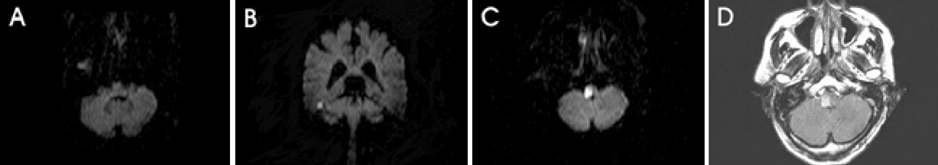
Figure 1 Case 1: A 69-year-old male patient with acute lateral medullary infarction. (A, B) At 8 h after onset of symptoms, initial diffusion-weighted imaging (DWI) shows no abnormal signal intensity. (C, D) At 63 h after onset of symptoms, the follow-up DWI and FLAIR imaging show a clear hyperintensity in the right lateral medullar area, matching the clinical presentation.
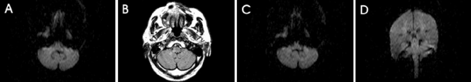
Figure 2 Case 22: A 67-year-old female patient with acute lateral medullary infarction. (A) At 6 h after onset of symptoms, initial DWI shows no abnormal signal intensity. (B) At 96 h after onset of symptoms, FLAIR imaging shows hyperintensity in the right lateral medullar area, matching the clinical presentation. In comparison, the follow-up DWI is considered normal (C, D).
Cited by 1 articles
-
Delayed Lesions on Diffusion-Weighted Imaging in Initially Lesion-Negative Stroke Patients
Kitae Kim, Beom Joon Kim, Jaewon Huh, Seong Kyu Yang, Mi Hwa Yang, Moon-Ku Han, Cheolkyu Jung, Byung Se Choi, Jae Hyoung Kim, Hee-Joon Bae
J Stroke. 2021;23(1):69-81. doi: 10.5853/jos.2020.02110.
Reference
-
1. Bogousslavsky J, Caplan L. Stroke syndromes. 2001. 2nd edn. New York: Cambridge University Press;534–536.2. Sacco RL, Freddo L, Bello JA, Odel JG, Onesti ST, Mohr JP. Wallenberg's lateral medullary syndrome. Clinicalmagnetic resonance imaging correlations. Arch Neurol. 1993. 50:609–614.3. Kistler JP, Buonanno FS, DeWitt LD, Davis KR, Brady TJ, Fisher CM. Vertebral-basilar posterior cerebral territory stroke - delineation by proton nuclear magnetic resonance imaging. Stroke. 1984. 15:417–426.
Article4. Isa K, Kimura K, Yasaka M, Minematsu K, Yamaguchi T. 23A case with frequent episodes of transient ischemic attack presenting the Wallenberg syndrome before and after the onset of brain infarction. Rinsho Shinkeigaku. 1999. 39:573–576.5. Kitis O, Calli C, Yunten N, Kocaman A, Sirin H. Wallenberg's lateral medullary syndrome: diffusionweighted imaging findings. Acta Radiol. 2004. 45:78–84.
Article6. Lövblad KO, Laubach HJ, Baird AE, Curtin F, Schlaug G, Edelman RR, et al. Clinical experience with diffusionweighted MR in patients with acute stroke. Am J Neuroradiol. 1998. 19:1061–1066.7. Oppenheim C, Stanescu R, Dormont D, Crozier S, Marro B, Samson Y, et al. False-negative diffusion-weighted MR findings in acute ischemic stroke. Am J Neuroradiol. 2000. 21:1434–1440.8. Narisawa A, Shamoto H, Shimizu H, Tominaga T, Yoshimoto T. Diffusion-weighted magnetic resonance imaging (MRI) in acute brain stem infarction. No To Shinkei. 2001. 53:1021–1026.9. Pedraza S, Osuna MT, Davalos A, Teruel J, Vera-Sancho J, Inaraja L. False negative diffusion in acute ischemic stroke. Rev Neurol. 2002. 34:1127–1129.10. Hirose Y, Mokuno K. Features of MRI diffusion weighted image in early stage of lateral medullary infarction presenting Wallenberg syndrome. Rinsho Shinkeigaku. 1998. 38:157–160.11. Kim JS. Pure lateral medullary infarction: clinical-radiological correlation of 130 acute, consecutive patients. Brain. 2003. 126:1864–1872.
Article12. Wang W, Goldstein S, Scheuer ML, Branstetter BF. Acute stroke syndrome with fixed neurological deficit and false-negative diffusion-weighted imaging. J Neuroimaging. 2003. 13:158–161.
Article13. Wang PY, Barker PB, Wityk RJ, Ulug AM, van Zijl PC, Beauchamp NJ Jr. Diffusion-negative stroke: a report of two cases. Am J Neuroradiol. 1999. 20:1876–1880.14. Sharbrough FW, Messick JM Jr, Sundt TM Jr. Correlation of continuous electroencephalograms with cerebral blood flow measurements during carotid endarterectomy. Stroke. 1973. 4:674–683.
Article15. Ginsberg M. The new language of cerebral ischemia. Am J Neuroradiol. 1997. 18:1435–1445.16. Lövblad KO, Jakob PM, Chen Q, Baird AE, Schlaug G, Warash S, et al. Turbo spin-echo diffusion-weighted MR of ischemic stroke. AJNR Am J Neuroradiol. 1998. 19:201–208.
- Full Text Links
-
- Actions
-
Cited
- CITED
-
- Close
- Share
-
- Similar articles
-
- Bilateral Medial Medullary Infarction Demonstrated by Diffusion-Weighted Imaging: Case Report
- Reversal of a Large Ischemic Lesion with Low Apparent Diffusion Coefficient Value by Rapid Spontaneous Recanalization
- A Case of Posterior Inferior Cerebellar Artery Infarction Presenting with Sudden Hearing Loss and Vertigo
- Lateral medullary infarction in a patient with Moyamoya disease associated with RNF213 variants: a case report
- A Case of Ipsilateral Hemiparesis in Lateral Medullary Infarction: As a Warning Sign of Acute Progression of Vertebral Artery Thrombosis
