Mild Encephalopathy with Reversible Lesion in the Splenium of the Corpus Callosum and Bilateral Frontal White Matter
- Affiliations
-
- 1Departments of Neurology and Radiology, Seoul Veterans Hospital, Seoul, Korea. hippocam@naver.com
- KMID: 1700688
- DOI: http://doi.org/10.3988/jcn.2007.3.1.53
Abstract
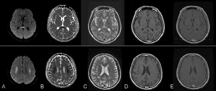
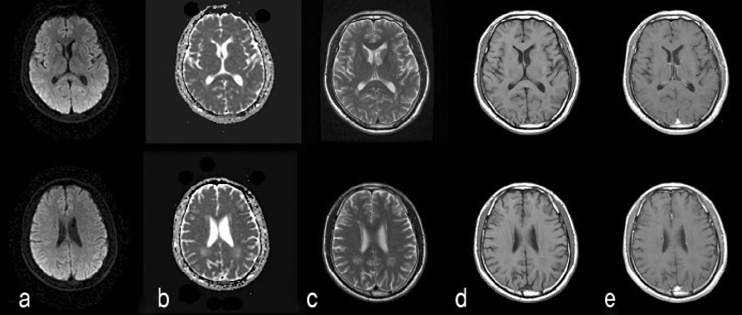
- A 59-year-old man visited an emergency room due to the sudden onset of severe dysarthria with a drowsy mental status. MRI demonstrated T2 prolongation and restricted diffusion involving the splenium of the corpus callosum and bilateral frontal white matter neurological signs and symptoms were mild, and the recovery was complete within a week. Follow-up MRI performed one month later revealed complete resolution of the lesions. The clinical and radiological courses were consistent with previously reported reversible isolated splenial lesions in mild encephalitis/encephalopathy except for the presence of frontal lesions. This case suggests that such reversible lesions can occur outside the splenium.
MeSH Terms
Figure
Cited by 4 articles
-
Reversible Splenium Lesion of the Corpus Callosum in Hemorrhagic Fever with Renal Failure Syndrome
Shin-Hye Baek, Dong-Ick Shin, Hyung-Suk Lee, Sung-Hyun Lee, Hye-Young Kim, Kyeong Seob Shin, Seung Young Lee, Ho-Seong Han, Hyun Jeong Han, Sang-Soo Lee
J Korean Med Sci. 2010;25(8):1244-1246. doi: 10.3346/jkms.2010.25.8.1244.Lateralization of Hypoglycemic Encephalopathy: Evidence of a Mechanism of Selective Vulnerability
Seung-Hwan Lee, Chang Don Kang, Sam Soo Kim, Woo-Suk Tae, Seo-Young Lee, Sung Hun Kim, Sung Hye Koh
J Clin Neurol. 2010;6(2):104-108. doi: 10.3988/jcn.2010.6.2.104.A Case of Scrub Typhus Related Encephalopathy Presenting as Rapidly Progressive Dementia
Jeong Hoon Park, Jae-Won Jang, Seung-Hwan Lee, Won Sup Oh, Sam Soo Kim
Dement Neurocogn Disord. 2017;16(3):83-86. doi: 10.12779/dnd.2017.16.3.83.Transient splenial lesions of the corpus callosum and infectious diseases
Kyu Sun Yum, Dong-Ick Shin
Acute Crit Care. 2022;37(3):269-275. doi: 10.4266/acc.2022.00864.
Reference
-
1. Takanashi J, Barkovich AJ, Shiihara T, Tada H, Kawatani M, Tsukafara H, et al. Widening spectrum of a reversible splenial lesion with transiently reduced diffusion. AJNR Am J Neuroradiol. 2006. 27:836–838.2. Takanashi J, Hirasawa K, Tada H. Reversible restricted diffusion of entire corpus callosum. J Neurol Sci. 2006. 247:101–104.
Article3. Tada H, Takanashi AJ, Barkovich H, Oba H, Maeda M, Tsukahara H, et al. Clinically mild encephalitis/encephalopathy with a reversible splenial lesion. Neurology. 2004. 63:1854–1858.
Article4. Seo HJ, Kim SY, Kim WM, Hong YJ, Sohn JH, Lee SM, et al. A case of encephalitis with a reversible splenial lesion on a diffusion weighted MRI image. J Korean Neurol Assoc. 2006. 24:507–510.5. Uchino A, Takasa Y, Nomiyama K, Egashira R, Kudo S. Acquired lesions of the corpus callosum: MR imaging. Eur Radiol. 2006. 16:905–914.
Article6. Hong JM, Park MS, Jun DC. Transient splenial lesion of the corpus callosum in patients with infectious disease. J Korean Neurol Assoc. 2005. 23:667–669.7. Kobata R, Tsukahara H, Nakai A, Tanizawa A, Ishimori Y, Kawamura Y, et al. Transient MR signal changes in the splenium of the corpus callosum in rotavirus encephalopathy: value of diffusion-weighted imaging. J Comput Assist Tomogr. 2002. 26:825–828.
Article8. Shin YE, Cho YW, Kim HA, Sohn SI, Lee HL, Lim JG, et al. Two cases of transient focal lesion in the splenium of the corpus callosum after aggravated seizures. J Korean Neurol Assoc. 2005. 23:111–1139.9. Takanashi J, Maeda M, Hayashi M. Neonate showing a reversible splenial lesion. Arch Neurol. 2005. 62:1481–1482.
- Full Text Links
- Actions
-
Cited
- CITED
-
- Close
- Share
- Similar articles
-
- Reversible Lesion in The splenium of The Corpus Callosum Induced by Topiramate in a Patient with Migraine
- A Case of Transient Isolated Splenial Lesion of the Corpus Callosum After New Onset Seizure
- Recurrent Clinically Mild Encephalitis/Encephalopathy with a Reversible Splenial Lesion (MERS) on Diffusion Weighted Imaging: A Case Report
- Reversible Splenial Lesion in the Corpus Callosum on MRI after Ingestion of a Herbicide Containing Glufosinate Ammonium: A Case Report
- Novel Information on Anatomic Factors Causing Grasp Reflex in Frontal Lobe Infarction: A Case Report


