A Case of Bronchiolitis Obliterans Organizing Pneumonia from Epstein-Barr Virus
- Affiliations
-
- 1Department of Internal Medicine, Yonsei University College of Medicine, Seoul, Korea. kom2d@netsgo.com
- 2Department of Internal Medicine and Pathology, National Health Iinsurance Corporation Ilsan Hospital, Koyang, Korea.
- 3Department of Radiology, National Health Iinsurance Corporation Ilsan Hospital, Koyang, Korea.
- KMID: 1518489
- DOI: http://doi.org/10.4046/trd.2007.62.1.51
Abstract
- In the average adult with a normal immune state, Epstein-Barr virus pneumonia is very rare, especially in the form of interstitial lung disease. According to recent studies, the Epstein-Barr virus is also associated with lymphocytic interstitial pneumonia, AIDS and Langerhans cell histiocytosis, but not with sarcoidosis. BOOP is caused by lung injury due to an infection or drug intoxication, and is related to connective tissue disease or bone marrow transplantation, but is sometimes idiopathic. We experienced a patient with symptoms and signs of interstitial lung disease, with confirmed BOOP and EBV ingection from an open lung biopsy and serologic examination, respectively Herein, this case is reported, with a review of the literature.
MeSH Terms
Figure
-

Figure 1 On H-E section, loose branching fibroblastic proliferations are observed within the terminal airway and alveolar spaces (A, H-E, ×200) with relatively mild interstitial inflammatory cell infiltration (B, H-E, ×400).

Figure 2 Chest radiograph shows diffuse ground glass opacity and reticular opacity in the both lungs (A). Chest PA after 5 months shows resolution of diffuse ground glass and reticular opacity in the both lungs (B).
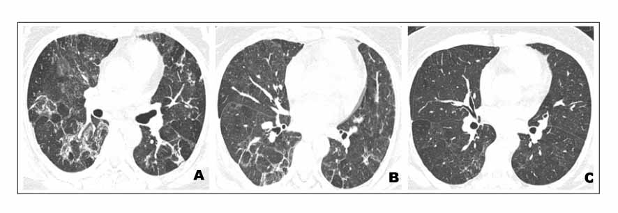
Figure 3 Axial CT image shows diffuse patch distributed ground glass opacity and consolidation along the bronchovascular bundles (A). Three week follow-up Chest CT shows improvement of ground glass opacity and consolidation in the both lungs (B). Follow-up Chest CT after 1 year demonstrates resolution of diffuse pulmonary infiltration. Mild ground glass opacity and reticular opacity are remained (C).
Cited by 1 articles
-
A Case of Bronchiolitis Obliterans Organizing Pneumonia Associated with Pandemic Influenza (H1N1 2009)
Min Hee Lim, Sang Taek Heo, Ho Cheol Kim, In-Gyu Bae, Jae hee Kim, In-Suk Kim, Sunjoo Kim, Gyung Hyuck Ko
Infect Chemother. 2010;42(2):112-116. doi: 10.3947/ic.2010.42.2.112.
Reference
-
1. Kim KS, Lee YM, Choi YS, Shin JH, Han GJ, Moon SH, et al. 2 case of idiopathic BOOP associated with rare radiologic finding. Tuberc Respir Dis. 1996. 43:228–235.2. Dodd JD, Muller NL. Bronchiolitis obliterans organizing pneumonia after bone marrow transplantation. J Comput Assist Tomogr. 2005. 29:540–543.3. Ahn MS, Koh YM, Shin J, Jeong HB, Lee SE, Chung YT. A case of Pneumoncystis carinii pneumonia with histopathologic finding of bronchiolitis obliterans organizing pneumonia in patient with AIDS. Tuberc Respir Dis. 1998. 45:444–450.4. Verma N, Soans B. Cryptogenic organizing pneumonia associated with Pneumocystis carinii infection and sirolimus therapy in a renal transplant patient. Australas Radiol. 2006. 50:68–70.5. Chantraumat C, Sittipunt C. Bronchiolitis obliterans organizing pneumonia caused by capsule-deficient cryptococcosis. Southeast Asian J Trop Med Public Health. 2005. 36:174–177.6. Epler GR. Bronchiolitis obliterans organizing pneumonia. Arch Intern Med. 2001. 161:158–164.7. Marzouk K, Corate L, Saleh S, Sharma OP. Epstein-Barr-virus-induced interstitial lung disease. Curr Opin Pulm Med. 2005. 11:456–460.8. Costabel U, Teschler H, Schoenfeld B, Hartung W, Nusch A, Guzman J, et al. BOOP in Europe. Chest. 1992. 102:Suppl. 14S–20S.
- Full Text Links
-
- Actions
-
Cited
- CITED
-
- Close
- Share
-
- Similar articles
-
- A case of idiopathic bronchiolitis obliterans organizing pneumonia
- A case of bronchiolitis obliterans organizing pneumonia associated with wheezing
- Dermatomyositis without elevation of creatine kinase presented as bronchiolitis obliterans organizing pneumonia
- Diffuse Micronodular Pattern of Bronchiolitis Obliterans Organizing Pneumonia: A Case Report
- Two Cases of Bronchiolitis Obliterans Organizing Pneumonia treated with Steroid and Cyclosporine therapy
