Korean J Gastroenterol.
2013 May;61(5):265-269. 10.4166/kjg.2013.61.5.265.
Ischemic Necrosis of the Cecum: A Single Center Experience
- Affiliations
-
- 1Department of General Surgery, Meram Medical Faculty, Necmettin Erbakan University, Konya, Turkey. ebubekir82@hotmail.com
- KMID: 1501648
- DOI: http://doi.org/10.4166/kjg.2013.61.5.265
Abstract
- BACKGROUND/AIMS
Isolated cecal necrosis is a rare cause of the surgical abdomen. Its manifestation is similar to that of acute appendicitis. Thirteen cases, who were pre-diagnosed with acute abdomen and were finally diagnosed with isolated cecal necrosis after operation have been evaluated alongside with literature.
METHODS
The records of 13 patients, who had isolated cecal necroses between 1995 and 2011 at Necmettin Erbakan University Meram Medical School's General Surgery Clinic (Turkey), were retrospectively evaluated.
RESULTS
Eight of the patients were male, whereas 5 were female. Their mean age was 68.0+/-11.7 (range 51-84) years. All the patients had at least one accompanying disease the most frequent of which were heart failure and chronic renal failure. Ten patients had right hemicolectomy and ileotransversostomy, two had right hemicolectomy and ileostomy, and one had wedge resection to the cecum by the help of linear stapler. Mortality was seen in 5 patients (38%) in the early postoperative period.
CONCLUSIONS
Isolated cecal necrosis should be considered in elderly patients with chronic diseases presenting with sudden right lower quadrant pains in the differential diagnosis. Isolated cecal necrosis may have a bad prognosis since it is seen in elderly patients with accompanying problems. Therefore, early diagnosis and immediate surgical management if necessary is important to reduce the risk of morbidity and mortality.
Keyword
MeSH Terms
Figure
-
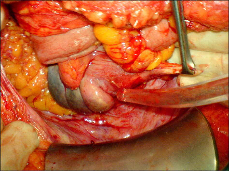
Fig. 1. Ischemic field (3×5 cm) on the cecal lateral wall.
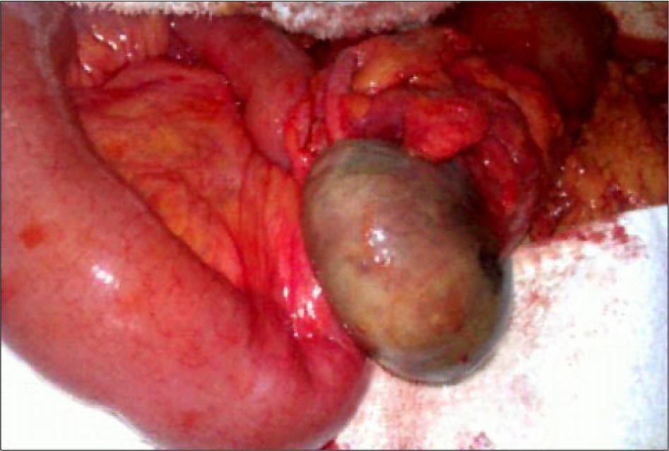
Fig. 2. Ischemic outlook covering almost the entire cecum.
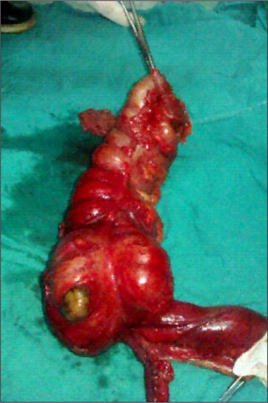
Fig. 4. (A) Ischemic field (2×2 cm) on the cecal anterior wall. (B) Cecal wedge resection material by the help of stapler. (C) The outlook of the cecum following resection with stapler.

Fig. 3. Right hemicolectomy was performed because of cecal necrosis.
Reference
-
References
1. Beck DE, de Aguilar-Nascimento JE. Surgical management and outcome in acute ischemic colitis. Ochsner J. 2011; 11:282–285.2. Yasuhara H. Acute mesenteric ischemia: the challenge of gastroenterology. Surg Today. 2005; 35:185–195.
Article3. Dirican A, Unal B, Bassulu N, Tatlı F, Aydin C, Kayaalp C. Isolated cecal necrosis mimicking acute appendicitis: a case series. J Med Case Rep. 2009; 3:7443–7447.
Article4. Borra S, Kleinfield M. Ischemic necrosis of the cecum: Findings in three dialysis patients. Geriatr Nephrol Urol. 1995; 5:15–19.
Article5. Kiyak G, Ozgün Y, Sarikaya SM, Korukluoğ lu B. Isolated cecal necrosis mimicking acute appendicitis. Turk J Gastroenterol. 2008; 19:71–72.6. Wiesner W, Mortelé KJ, Glickman JN, Ros PR. "Cecal gangrene": a rare cause of right-sided inferior abdominal quadrant pain, fever, and leukocytosis. Emerg Radiol. 2002; 9:292–295.
Article7. Rist CB, Watts JC, Lucas RJ. Isolated ischemic necrosis of the cecum in patients with chronic heart disease. Dis Colon Rectum. 1984; 27:548–551.
Article8. Hargrove WC 3rd, Rosato EF, Hicks RE, Mullen JL. Cecal necrosis after open-heart operation. Ann Thorac Surg. 1978; 25:71–73.
Article9. Schuler JG, Hudlin MM. Cecal necrosis: infrequent variant of ischemic colitis. Report of five cases. Dis Colon Rectum. 2000; 43:708–712.10. Simon AM, Birnbaum BA, Jacobs JE. Isolated infarction of the cecum: CT findings in two patients. Radiology. 2000; 214:513–516.
Article11. Friedell ML. Cecal necrosis in the dialysis-dependent patient. Am Surg. 1985; 51:621–622.12. Margovsky A, Grieve DA. Cocaine-induced isolated caecum necrosis. ANZ J Surg. 2001; 71:321–322.
Article13. Perko Z, Bilan K, Vilović K, et al. Partial cecal necrosis treated by laparoscopic partial cecal resection. Coll Antropol. 2006; 30:937–939.14. Hoeffel C, Crema MD, Belkacem A, et al. Multidetector row CT: spectrum of diseases involving the ileocecal area. Radiographics. 2006; 26:1373–1390.
Article
