Korean J Ophthalmol.
2012 Dec;26(6):473-477. 10.3341/kjo.2012.26.6.473.
Optic Disc Atrophy in Patient with Posner-Schlossman Syndrome
- Affiliations
-
- 1Department of Ophthalmology, Samsung Medical Center, Sungkyunkwan University School of Medicine, Seoul, Korea. cwkee@skku.edu
- 2Department of Ophthalmology, Busan Paik Hospital, Inje University School of Medicine, Busan, Korea.
- KMID: 1499689
- DOI: http://doi.org/10.3341/kjo.2012.26.6.473
Abstract
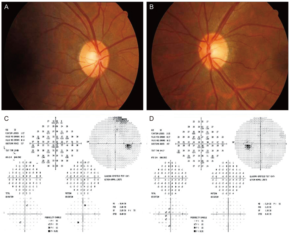
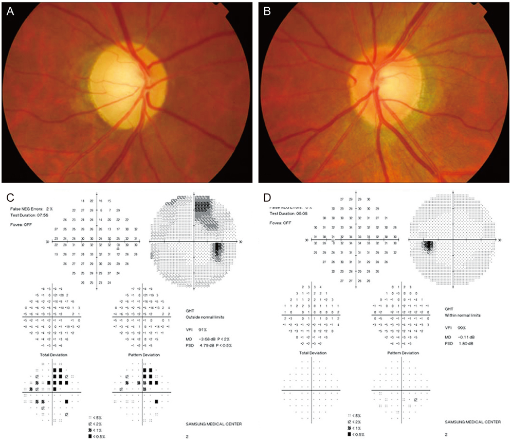
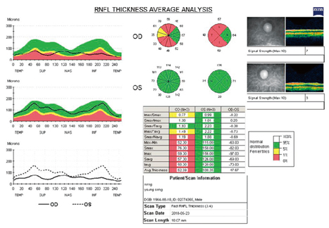
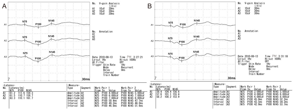
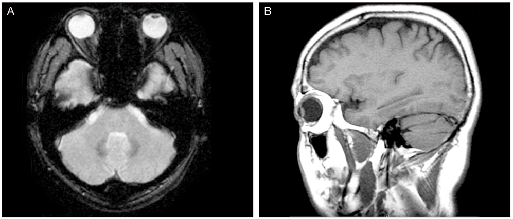
- A 32-year-old man with blurred vision in the right eye and headache presented with anterior uveitis, an intraocular pressure (IOP) of 60 mmHg, an open angle, no visual field defects, and normal optic nerve. He had a history of five previous similar attacks. In each of the previous instances, his anterior uveitis and high IOP were controlled with antiglaucoma medications and topical steroids. However, at the fifth attack, his optic disc was pale and a superior paracentral visual field defect was shown. Brain magnetic resonance image studies were normal. This case represents that a recurrent Posner-Schlossman syndrome (PSS)-induced optic disc atrophy likely due to ocular ischemia caused by a recurrent, high IOP. Although PSS is a self-limiting syndrome, we should manage high IOP and prevent ischemia of the optic nerve head by treating with ocular antihypertensive medications.
MeSH Terms
Figure
Cited by 2 articles
-
Clinical Features and Risk Factors of Glaucomatous Change in Posner-Schlossman Syndrome
Eun Jung Lee, Young Kyo Kwun, Dong Hoon Shin, Chang Won Kee
J Korean Ophthalmol Soc. 2015;56(6):938-943. doi: 10.3341/jkos.2015.56.6.938.Point-of-care monitoring of perioperative intraocular pressure using portable tonometry in a patient with Posner-Schlossman syndrome: a case report
Sung-Hoon Kim, Jin-Ho Rhim, Young-Jin Moon, Jihion Yu, Jong-Yeon Park, Ashish Bangaari
Korean J Anesthesiol. 2014;66(3):248-251. doi: 10.4097/kjae.2014.66.3.248.
Reference
-
1. Posner A, Schlossman A. Syndrome of unilateral recurrent attacks of glaucoma with cyclitic symptoms. Arch Ophthal. 1948. 39:517–535.2. Raitta C, Vannas A. Glaucomatocyclitic crisis. Arch Ophthalmol. 1977. 95:608–612.3. Kass MA, Becker B, Kolker AE. Glaucomatocyclitic crisis and primary open-angle glaucoma. Am J Ophthalmol. 1973. 75:668–673.4. Kim R, Van Stavern G, Juzych M. Nonarteritic anterior ischemic optic neuropathy associated with acute glaucoma secondary to Posner-Schlossman syndrome. Arch Ophthalmol. 2003. 121:127–128.5. Irak I, Katz BJ, Zabriskie NA, Zimmerman PL. Posner-Schlossman syndrome and nonarteritic anterior ischemic optic neuropathy. J Neuroophthalmol. 2003. 23:264–267.6. Hirose S, Ohno S, Matsuda H. HLA-Bw54 and glaucomatocyclitic crisis. Arch Ophthalmol. 1985. 103:1837–1839.7. Teoh SB, Thean L, Koay E. Cytomegalovirus in aetiology of Posner-Schlossman syndrome: evidence from quantitative polymerase chain reaction. Eye (Lond). 2005. 19:1338–1340.8. Yamamoto S, Pavan-Langston D, Tada R, et al. Possible role of herpes simplex virus in the origin of Posner-Schlossman syndrome. Am J Ophthalmol. 1995. 119:796–798.9. Hayreh SS. Anterior ischemic optic neuropathy. Arch Neurol. 1981. 38:675–678.
- Full Text Links
- Actions
-
Cited
- CITED
-
- Close
- Share
- Similar articles
-
- A Case of Posner-Schlossman Syndrome with Retinal Arterial Tortuosity in a Young Male
- Clinical Factors of Glaucomatous Change in Patients with Posner-Schlossman Syndrome
- Point-of-care monitoring of perioperative intraocular pressure using portable tonometry in a patient with Posner-Schlossman syndrome: a case report
- A Case of Posner-Schlossman Syndrome following after Strabismus Surgery
- A Case of Foster Kennedy Syndrome

