Pre- and Post-Angioplasty Perfusion CT with Acetazolamide Challenge in Patients with Unilateral Cerebrovascular Stenotic Disease
- Affiliations
-
- 1Department of Neurosurgery, Gangneung Asan Hospital, University of Ulsan College of Medicine, Gangneung, Korea.
- 2Department of Radiology, Gangneung Asan Hospital, University of Ulsan College of Medicine, Gangneung, Korea.
- 3Department of Neurology, Gangneung Asan Hospital, University of Ulsan College of Medicine, Gangneung, Korea.
- 4Department of Preventive Medicine, College of Medicine, Gwandong University, Gangneung, Korea. wspark@kd.ac.kr
- KMID: 1499332
- DOI: http://doi.org/10.3340/jkns.2013.54.4.280
Abstract
OBJECTIVE
Perfusion computed tomography (PCT) has the ability to measure quantitative value and produce maps of mean transit time (MTT), cerebral blood flow (CBF), and cerebral blood volume (CBV). We assessed cerebral hemodynamics by using these parameters and acetazolamide (ACZ) challenge for pre- and post-procedural evaluation in patients with unilateral cerebrovascular stenotic disease.
METHODS
Thirty patients underwent pre-procedural PCT with ACZ challenge, and 24 patients (80%) was conducted follow up PCT after angioplasty with same protocol. The mean MTT, CBF, and CBV were measured and compared in both middle cerebral arterial (MCA) territories before and after ACZ challenge. Hemispheric ratio and percent change after ACZ challenge were calculated before and after angioplasty.
RESULTS
The mean stenosis rate was 76.6%. Significant increases in MTT (32.6%, p=0.000) and significant decreases in CBF (-14.2%, p=0.000) were found in stenotic side MCA territories. After ACZ challenge, there were significant changes in MTT (37.4%, p=0.000), CBF (-13.1%, p=0.000), and CBV (-10.5%, p=0.001) in pre-procedural perfusion study. However, no significant increases were found in MTT, or decreases in CBF and CBV in post-procedural study. There were no significant changes after ACZ challenge also. In addition, the degrees of these changes (before and after ACZ challenge) were highly correlated with the stenotic degrees in pre-procedural perfusion study.
CONCLUSION
PCT with ACZ challenge appears to be a useful tool to assess the cerebral perfusion status especially in patients with unilateral symptomatic stenotic disease.
Keyword
MeSH Terms
Figure
-
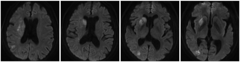
Fig. 1 Diffusion magnetic resonance imaging (MRI) of a 64-year-old female at admission, showing typical borderzone infarction on the right hemisphere. Perfusion computed tomography (PCT) and internal cerebral artery (ICA) angiogram of this patient are shown on Fig. 2 and 3 respectively.
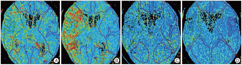
Fig. 2 Pre- and post-angioplasty mean transit time (MTT) maps before and after acetazolamide (ACZ) challenge. A : Pre-angioplasty MTT map before ACZ challenge. The hemispheric ratio of right middle cerebral artery (MCA) territory versus left MCA territory is 165.1%. B : Pre-angioplasty MTT map after ACZ challenge. The hemispheric ratio is 306.4%. C : Post-angioplasty MTT map before ACZ challenge. The hemispheric ratio of right MCA territory versus left MCA territory is 112.2%. D : Post-angioplasty MTT map after ACZ challenge. The hemispheric ratio is 117.3%.
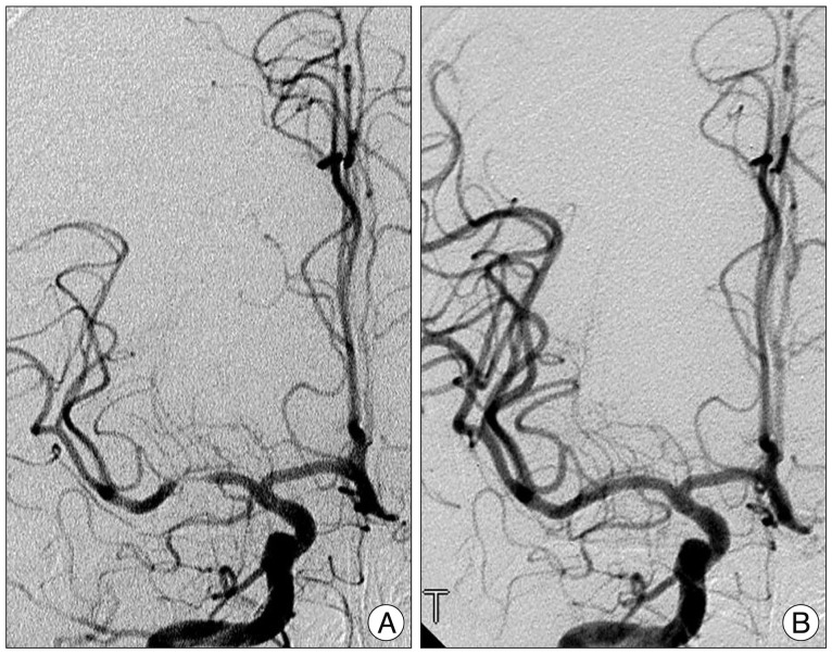
Fig. 3 Right internal cerebral artery (ICA) angiograms before (A) and after (B) stent-angioplasty. A : Right ICA angiogram shows severe stenosis of middle cerebral artery (MCA) up to 84%. B : The stenotic lesion was fully recovered after stent-angioplasty.
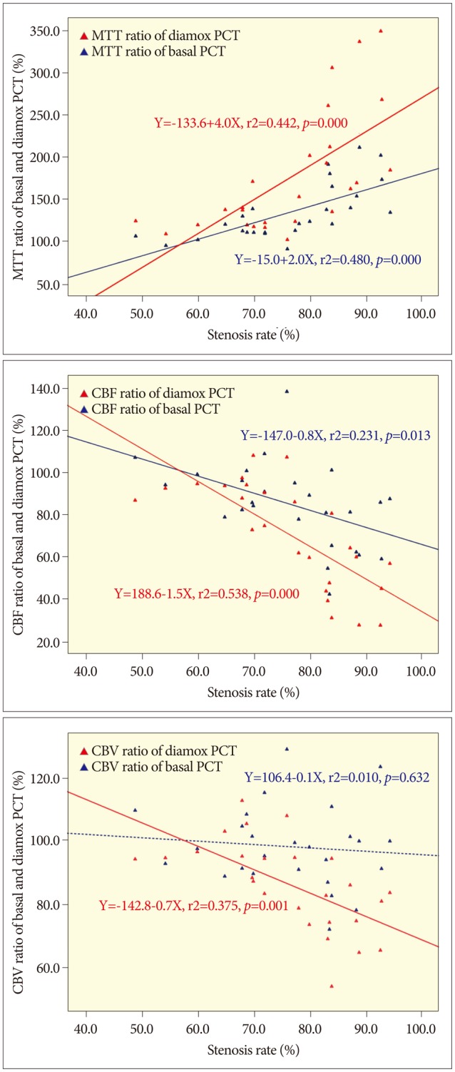
Fig. 4 Linear regression plots of each parameter before angioplasty. A : Mean transit time (MTT) ratio shows positive linear relationship with the stenosis rate before acetazolamide (ACZ) challenge (Basal MTT), and the gradient is steeper after acetazolamide challenge (Diamox MTT). B : Cerebral blood flow (CBF) ratio before ACZ challenge (Basal CBF) shows negative linear relationship, and the gradient is steeper after ACZ challenge (Diamox CBF). C : Cerebral blood volume (CBV) ratio before ACZ challenge (Basal CBV) does not show significant linear relationship, however it shows significant negative relationship after ACZ challenge (Diamox CBV). Blue dot and line : values before ACZ challenge. Red dot and line: values after ACZ challenge.

Fig. 5 Linear regression plots of each parameter's hemispheric ratio change {absolute value of ratio difference=[hemispheric ratio after acetazolamide (ACZ) challenge-hemispheric ratio before ACZ challenge]} versus stenosis rate. The degrees of all the three parameters' ratio changes show positive linear relationships with the stenosis rate. The horizontal black dotted line indicates zero percent of hemispheric ratio change, and all the three lines cross with the horizontal black dotted line at near 60& of stenosis rate, and continue to rise. The gradient of mean transit time (MTT) is steeper than the others. Red dot and line : MTT. Green dot and line : cerebral blood flow (CBF). Blue dot and line: cerebral blood volume (CBV).
Reference
-
1. Ahn JY, Lee BH, Choi EW, Kim OJ, Choi BO. Improved cerebral perfusion after stent-assisted angioplasty for middle cerebral artery stenosis. J Korean Neurosurg Soc. 2002; 31:364–368.2. Chen A, Shyr MH, Chen TY, Lai HY, Lin CC, Yen PS. Dynamic CT perfusion imaging with acetazolamide challenge for evaluation of patients with unilateral cerebrovascular steno-occlusive disease. AJNR Am J Neuroradiol. 2006; 27:1876–1881. PMID: 17032859.3. Chimowitz MI, Lynn MJ, Derdeyn CP, Turan TN, Fiorella D, Lane BF, et al. Stenting versus aggressive medical therapy for intracranial arterial stenosis. N Engl J Med. 2011; 365:993–1003. PMID: 21899409.4. Cianfoni A, Colosimo C, Basile M, Wintermark M, Bonomo L. Brain perfusion CT : principles, technique and clinical applications. Radiol med. 2007; 112:1225–1243. PMID: 18074193.
Article5. Eastwood JD, Alexander MJ, Petrella JR, Provenzale JM. Dynamic CT perfusion imaging with acetazolamide challenge for the preprocedural evaluation of a patient with symptomatic middle cerebral artery occlusive disease. AJNR Am J Neuroradiol. 2002; 23:285–287. PMID: 11847056.6. Fiorella D, Derdeyn CP, Lynn MJ, Barnwell SL, Hoh BL, Levy EI, et al. Detailed analysis of periprocedural strokes in patients undergoing intracranial stenting in stenting and aggressive medical management for preventing recurrent stroke in intracranial stenosis (SAMMPRIS). Stroke. 2012; 43:2682–2688. PMID: 22984008.
Article7. Furukawa M, Kashiwagi S, Matsunaga N, Suzuki M, Kishimoto K, Shirao S. Evaluation of cerebral perfusion parameters measured by perfusion CT in chronic cerebral ischemia : comparison with xenon CT. J Comput Assist Tomogr. 2002; 26:272–278. PMID: 11884786.
Article8. Koenig M, Kraus M, Theek C, Klotz E, Gehlen W, Heuser L. Quantitative assessment of the ischemic brain by means of perfusion-related parameters derived from perfusion CT. Stroke. 2001; 32:431–437. PMID: 11157178.
Article9. Lee YH, Kwon W, Lee MS, Huh YM, Kim MS. Development of PC-based software to analyze dynamic cerebral perfusion CT quantitatively and to reformat perfusion maps. J Korean Radiol Soc. 2005; 53:79–84.
Article10. Lee TJ, Lee MS, Kim MS, Hong IS, Lee YH, Lee JY, et al. The utility of first-pass perfusion CT in hyperacute ischemic stroke : early experience. J Korean Radiol Soc. 2003; 49:231–235.
Article11. Levy EI, Hanel RA, Boulos AS, Bendok BR, Kim SH, Gibbons KJ, et al. Comparison of periprocedure complications resulting from direct stent placement compared with those due to conventional and staged stent placement in the basilar artery. J Neurosurg. 2003; 99:653–660. PMID: 14567599.
Article12. Marchal G, Beaudouin V, Rioux P, de la Sayette V, Le Doze F, Viader F, et al. Prolonged persistence of substantial volumes of potentially viable brain tissue after stroke : a correlative PET-CT study with voxel-based data analysis. Stroke. 1996; 27:599–606. PMID: 8614914.
Article13. Mayer TE, Hamann GF, Baranczyk J, Rosengarten B, Klotz E, Wiesmann M, et al. Dynamic CT perfusion imaging of acute stroke. AJNR Am J Neuroradiol. 2000; 21:1441–1449. PMID: 11003276.14. Nabavi DG, Cenic A, Craen RA, Gelb AW, Bennett JD, Kozak R, et al. CT assessment of cerebral perfusion : experimental validation and initial clinical experience. Radiology. 1999; 213:141–149. PMID: 10540654.
Article15. Nariai T, Suzuki R, Hirakawa K, Maehara T, Ishii K, Senda M. Vascular reserve in chronic cerebral ischemia measured by the acetazolamide challenge test : comparison with positron emission tomography. AJNR Am J Neuroradiol. 1995; 16:563–570. PMID: 7793382.16. Park JC, Kim JE, Kang HS, Sohn CH, Lee DS, Oh CW, et al. CT perfusion with angiography as a substitute for both conventional digital subtraction angiography and acetazolamide-challenged SPECT in the follow-up of postbypass patients. Cerebrovasc Dis. 2010; 30:547–555. PMID: 20948198.
Article17. Reichenbach JR, Röther J, Jonetz-Mentzel L, Herzau M, Fiala A, Weiller C, et al. Acute stroke evaluated by time-to-peak mapping during initial and early follow-up perfusion CT studies. AJNR Am J Neuroradiol. 1999; 20:1842–1850. PMID: 10588107.18. Röther J, Jonetz-Mentzel L, Fiala A, Reichenbach JR, Herzau M, Kaiser WA, et al. Hemodynamic assessment of acute stroke using dynamic single-slice computed tomographic perfusion imaging. Arch Neurol. 2000; 57:1161–1166. PMID: 10927796.
Article19. Shi M, Wang S, Zhou H, Cheng Y, Feng J, Wu J. Wingspan stenting of symptomatic middle cerebral artery stenosis and perioperative evaluation using high-resolution 3 Tesla MRI. J Clin Neurosci. 2012; 19:912–914. PMID: 22341146.
Article20. Siddiq F, Chaudhry SA, Khatri R, Rodriguez GJ, Tummala R, Suri MF, et al. Rate of postprocedural stroke and death in SAMMPRIS trial-eligible patients treated with intracranial angioplasty and/or stent placement in practice. Neurosurgery. 2012; 71:68–73. PMID: 22382209.
Article21. Tomandl BF, Klotz E, Handschu R, Stemper B, Reinhardt F, Huk WJ, et al. Comprehensive imaging of ischemic stroke with multisection CT. Radiographics. 2003; 23:565–592. PMID: 12740462.
Article22. Trojanowska A, Drop A, Jargiello T, Wojczal J, Szczerbo-Trojanowska M. Changes in cerebral hemodynamics after carotid stenting : evaluation with CT perfusion studies. J Neuroradiol. 2006; 33:169–174. PMID: 16840959.
Article23. Waaijer A, van der Schaaf IC, Velthuis BK, Quist M, van Osch MJ, Vonken EP, et al. Reproducibility of quantitative CT brain perfusion measurements in patients with symptomatic unilateral carotid artery stenosis. AJNR Am J Neuroradiol. 2007; 28:927–932. PMID: 17494672.24. Wintermark M, Reichhart M, Thiran JP, Maeder P, Chalaron M, Schnyder P, et al. Prognostic accuracy of cerebral blood flow measurement by perfusion computed tomography, at the time of emergency room admission, in acute stroke patients. Ann Neurol. 2002; 51:417–432. PMID: 11921048.
Article25. Wintermark M, Thiran JP, Maeder P, Schnyder P, Meuli R. Simultaneous measurement of regional cerebral blood flow by perfusion CT and stable xenon CT : a validation study. AJNR Am J Neuroradiol. 2001; 22:905–914. PMID: 11337336.
- Full Text Links
-
- Actions
-
Cited
- CITED
-
- Close
- Share
-
- Similar articles
-
- Utility of Acetazolamide-challenged CT Perfusion in Patients with High-grade Carotid Stenosis
- Acetazolamide-Challenged Brain CT Perfusion before and after Carotid Stenting
- Prediction of Prognosis by Acetazolamide Brain Perfusion SPECT in Patients with Arteriovenous Malformation
- Useful parameters utilizing perfusion CT study to evaluate the hemodynamic status in chronic ischemic stroke patients
- Diamox-enhanced Brain SPECT in Cerebrovascular Diseases
