Idiopathic Lenticulostriate Artery Pseudoaneurysm Protruding into the Lateral Ventricle: A Case Report
- Affiliations
-
- 1Department of Neurosurgery, Seoul National University Bundang Hospital, Seongnam, Korea. bang78425@hanmail.net
- 2Department of Pathology, Seoul National University Bundang Hospital, Seongnam, Korea.
- KMID: 1491447
- DOI: http://doi.org/10.7461/jcen.2013.15.3.246
Abstract
- We report a rare case of an idiopathic pseudoaneurysm causing intraventricular hemorrhage (IVH). A 28-year-old man presented with sudden onset of severe headache. He underwent external ventricular drainage for an isolated IVH in the right lateral ventricle. Digital subtraction angiography (DSA) revealed that the aneurysm (7.5x4.5 mm) arose from the distal part of the medial lenticulostriate artery. Following removal of the external ventricular drainage catheter, the aneurysm decreased in size (4.0x2.3 mm). However, follow-up DSA revealed a slightly enlarged aneurysm (4.2x3.2 mm) with morphologic change. The aneurysm was clipped via the interhemispheric transcallosal approach, but postoperative DSA revealed a residual aneurysm sac beside the clips. Given the risk of rebleeding, a second operation was planned for complete resection of the aneurysm. After revised craniotomy and careful dissection of the caudate nucleus, the aneurysm sac was completely resected. Histopathological examination revealed that the aneurysm was a pseudoaneurysm. The patient recovered without any neurological sequel and was discharged. To the best of our knowledge, this is the first reported case of an idiopathic lenticulostriate artery pseudoaneurysm protruding into the right lateral ventricle and causing an IVH that was successfully treated with microsurgical resection.
MeSH Terms
Figure
-
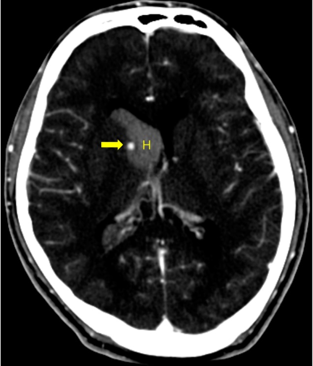
Fig. 1 Initial enhanced computed tomography images. Pure intraventricular hemorrhage is observed in the right lateral ventricle. Small nodular enhancing lesion is located beside head of caudate nucleus (Arrow: enhanced aneurysm, H: hematoma).

Fig. 2 Digital subtraction angiography (DSA) images. (A) Aneurysm from lenticulostriate artery is found in initial DSA. (Arrow: aneurysm, Arrowhead: lenticulostriate artery). (B) Follow-up DSA after initial clipping shows residual aneurysm with applied clips. (Arrow: aneurysm, Arrowhead: clips). (C) Final DSA shows disappeared aneurysm after resection.
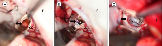
Fig. 3 Intraoperative photography. (A) Right lateral ventricle is exposed by interhemispheric approach. Saccular aneurysm sac is observed with small bleb. (Arrow: aneurysm, F: falx cerebri, CC: corpus callosum). (B) Clips are applied at the margin of caudate nucleus. (Arrow: clips, F: falx cerebri, CC: corpus callosum). (C) The head of right caudate nucleus is dissected and pseudoaneurysm is totally resected (Arrow: caudate nucleus, F: falx cerebri).
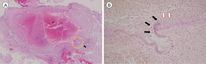
Fig. 4 Photomicrography. (A) Hematoxylin-eosin stain, original magnification ×40. (*: Recent thrombi with partial organization are observed, Arrow: partially ruptured vascular wall). (B) Elastin stain, original magnification ×200, this is magnification of yellow circle area in Fig. 4A (Black arrow: elastin layer is stained, White arrow: elastin layer is discontinued at the wall of hematoma).
Reference
-
1. Chapot R, Houdart E, Saint-Maurice JP, Aymard A, Mounayer C, Lot G, et al. Endovascular treatment of cerebral mycotic aneurysms. Radiology. 2002; 2. 222(2):389–396. PMID: 11818604.
Article2. Cohen JE, Gomori JM, Segal R, Spivak A, Margolin E, Sviri G, et al. Results of endovascular treatment of traumatic intracranial aneurysms. Neurosurgery. 2008; 9. 63(3):476–485. discussion 485-6. PMID: 18812959.
Article3. Inci S, Arat A, Ozgen T. Distal anterior choroidal artery aneurysms. Surg Neurol. 2007; 1. 67(1):46–52. discussion 52. PMID: 17210297.
Article4. Konishi Y, Kadowaki C, Hara M, Takeuchi K. Aneurysms associated with moyamoya disease. Neurosurgery. 1985; 4. 16(4):484–491. PMID: 3990927.
Article5. Madhugiri VS, Gundamaneni SK, Yadav AK, Sasidharan GM, Roopesh KV. Idiopathic intraventricular aneurysm presenting with intraventricular hemorrhage: Case report and review of the literature. Pediatr Neurosurg. 2012; 48(3):174–180. PMID: 23406825.
Article6. Medel R, Crowley RW, Hamilton DK, Dumont AS. Endovascular obliteration of an intracranial pseudoaneur ysm: the utility of Onyx. J Neurosurg Pediatr. 2009; 11. 4(5):445–448. PMID: 19877777.7. Miyake H, Ohta T, Kajimoto Y, Ogawa R, Deguchi J. Intraventricular aneurysms - Three case reports. Neurol Med Chir (Tokyo). 2000; 1. 40(1):55–60. PMID: 10721256.8. Saket RR, Hetts SW, Tatum JK, Glastonbury CM. CT and MRI findings of sphenoid sinus internal carotid artery pseudoaneurysm: An important and challenging differential diagnosis for a skull base mass. Clin Radiol. 2012; 8. 67(8):815–820. PMID: 22336670.
Article9. Sanli AM, Cekirge S, Sekerci Z. Aneurysm of the distal anterior cerebral artery radiologically mimicking a ventricular mass. J Neurosurg. 2011; 4. 114(4):1061–1064. PMID: 20635851.10. Ungersbock K, Perneczky A. Intraventricular aneurysm of the medial posterior choroid artery clipped via the contralateral transcallosal approach. Acta Neurochir (Wien). 1986; 82(1-2):24–27. PMID: 3751701.11. Urbach H, Meyer B, Cedzich C, Solymosi L. Posterior inferior cerebellar artery aneurysm in the fourth ventricle. Neuroradiology. 1995; 5. 37(4):267–269. PMID: 7666957.
Article
- Full Text Links
-
- Actions
-
Cited
- CITED
-
- Close
- Share
-
- Similar articles
-
- Spontaneous Resolution of Pulmonary Artery Pseudoaneurysm after Tube Thoracostomy
- Splenic Artery Pseudoaneurysm Complicating Chronic Pancreatitis: A Case Report
- Pseudoaneurysm of the Lateral Inferior Genicular Artery after Arthroscopic Partial Meniscectomy of Lateral Meniscus: A Case Report
- Glioblastoma Multiforme in the Trigone of the Lateral Ventricle: A Case Report
- Popliteal Artery Pseudoaneurysm after Arthroscopic Posterior Cruciate Ligament Reconstruction: A Case Report
