Alteration of Ventricular Repolarization by Intracoronary Infusion of Normal Saline in Patients With Variant Angina
- Affiliations
-
- 1Cardiovascular Center, Wonkwang University Hospital, Iksan, Korea. cardionh@wonkwang.ac.kr
- 2The Institute of Medical Sciences, Iksan, Korea.
- KMID: 1490697
- DOI: http://doi.org/10.4070/kcj.2009.39.6.223
Abstract
-
BACKGROUND AND OBJECTIVES: During coronary angiography and interventional procedures, catheters that are engaged in a coronary ostium are routinely flushed, typically with normal saline, to expel blood from the catheter or to inject a pharmacologic agent. Saline contains sodium and chloride ions. Such injections may affect the electrophysiologic properties of the myocardium; however, the effect of normal saline on ventricular repolarization has not been established in patients with variant angina.
SUBJECTS AND METHODS
We studied 51 consecutive patients with variant angina. Five mL of normal saline (NS) or 5% dextrose solution (DW) were infused into the left coronary artery in random order. We measured the heart rate, QT interval, and T-wave amplitude using Mac-Lac 5.2.
RESULTS
The baseline clinical characteristics were not different between the NS {n=30 (14 males); mean age, 56+/-10 years} and the 5% DW groups {n=21 (7 males); mean age, 59+/-10 years}. The changes in the mean corrected QT (QTc) interval were significantly increased at the time of infusion of NS compared to 5% DW (45.1+/-30.3 vs. 20.9+/-23.3 ms, p=0.004). There was a T-wave amplitude change >0.2 mV in at least one-lead in 27 patients (90.0%) during NS infusion compared to 7 patients (33.3%) during 5% DW infusions (p=0.001). No significant changes in heart rate and blood pressure were noted during of the infusions.
CONCLUSION
NS was associated with prolongation of ventricular repolarization in patients with variant angina.
MeSH Terms
Figure
-

Fig. 1 A representative measurement of the QT interval and T amplitude using Mac-Lab 5.2. The QT interval was 395 ms and T amplitude was 0.55 mV with a speed of 100 mm/sec in lead III.
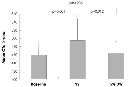
Fig. 2 Comparison of the mean QTc interval after intracoronary injection. NS: normal saline, DW: dextrose water, QTc: corrected QT interval by Bazett's formula.
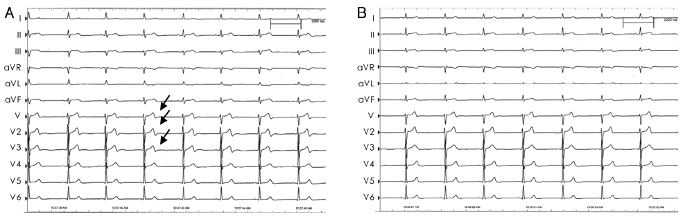
Fig. 3 ECGs recorded during intracoronary injection in a single patient. Note the marked changes in T-waves (arrow) and QT duration during and immediately after intracoronary normal saline injection (A). In contrast, T-waves and QT intervals remain stable during intracoronary 5% dextrose water injection (B). ECG: electrocardiogram.
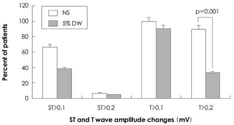
Fig. 4 Percentage of patients with ST and T-wave changes in at least one lead during and immediately after intracoronary injections. NS: normal saline, DW: dextrose water.
Reference
-
1. Dodek A, Boone JA, Hooper RO, Kavanagh-Gray D, Macdonald IL, Peretz DI. Complications of coronary arteriography. Can Med Assoc J. 1983. 128:934–936.2. Missri J, Jeresaty RM. Ventricular fibrillation during coronary angiography: reduced incidence with nonionic contrast media. Cathet Cardiovasc Diagn. 1990. 19:4–7.3. Wolf GL, Le Veen RF, Mulry C, Kilzer K. The influence of contrast media additives upon ventricular fibrillation thresholds during coronary angiography in ischemic and normal canine hearts. Cardiovasc Intervent Radiol. 1981. 4:145–147.4. Snyder CF, Formanek A, Frech RS, Amplatz K. The role of sodium in promoting ventricular arrhythmia during selective coronary arteriography. Am J Roentgenol Radium Ther Nucl Med. 1971. 113:567–571.5. Pedersen HK. Electrolyte addition to nonionic contrast media: cardiac effects during experimental coronary arteriography. Acta Radiol Suppl. 1996. 405:1–31.6. Bååth L, Besjakov J, Oksendal A. Sodium-calcium balance in nonionic contrast media: effects on the risk of ventricular fibrillation in the isolated rabbit heart. Invest Radiol. 1993. 28:223–227.7. Hillis LD, Braunwald E. Coronary-artery spasm. N Engl J Med. 1978. 299:695–702.8. Severi S, Davies G, Maseri A, Marzullo P, L'Abbate A. Long-term prognosis of "variant" angina with medical treatment. Am J Cardiol. 1980. 46:226–232.9. Kang JA, Lee YS, Jeong SH, et al. Clinical characteristics of patients with variant angina. Korean J Med. 2002. 63:195–202.10. Suzuki M, Nishizaki M, Arita M, et al. Increased QT dispersion in patients with vasospastic angina. Circulation. 1998. 98:435–440.11. Parchure N, Batchvarov V, Malik M, Camm AJ, Kaski JC. Increased QT dispersion in patients with Prinzmetal's variant angina and cardiac arrest. Cardiovasc Res. 2001. 50:379–385.12. Kim NH, Jeong JW, Song MJ, et al. The acute effects of cigarette smoking on the heterogeneity of ventricular repolarization in healthy subjects: the relationship between QT dispersion and coronary flow feserve. Korean Circ J. 2005. 35:448–453.13. De Ambroggi L, Negroni MS, Monza E, Bertoni T, Schwartz PJ. Dispersion of ventricular repolarization in the long QT syndrome. Am J Cardiol. 1991. 68:614–620.14. Dritsas A, Sbarouni E, Gilligan D, Nihoyannopoulos P, Oakley CM. QT-interval abnormalities in hypertrophic cardiomyopathy. Clin Cardiol. 1992. 15:739–742.15. Fei L, Goldman JH, Prasad K, et al. QT dispersion and RR variations on 12-lead ECGs in patients with congestive heart failure secondary to idiopathic dilated cardiomyopathy. Eur Heart J. 1996. 17:258–263.16. Kim NH, Cho JG, Lee SH, et al. Value of QT dispersion as a predictor of coronary artery stent restenosis. Korean Circ J. 2000. 30:555–562.17. Choi H. Dispersion of QT interval and other repolarization indexes in acute myocardial infarction. Korean Circ J. 1997. 27:1289–1297.18. Kuo CS, Reddy CP, Munakata K, Surawicz B. Mechanism of ventricular arrhythmias caused by increased dispersion of repolarization. Eur Heart J. 1985. 6:63–70.19. Surawicz B. The QT interval and cardiac arrhythmias. Annu Rev Med. 1987. 38:81–90.20. Bazett HC. An analysis of the time-relations of electrocardiograms. Heart. 1918. 7:353–370.21. Romanelli MF, Meissner MD, Fromm BS, Spears JR. Prominent ECG repolarization changes associated with intracoronary infusion of normal saline: comparisons with alternate coronary catheter flush solutions. Catheter Cardiovasc Interv. 1999. 48:359–364.22. Adams DF, Fraser DB, Abrams HL. The complications of coronary arteriography. Circulation. 1973. 48:609–618.23. Murdock DK, Euler DE, Becker DM, Murdock JD, Scanlon PJ, Gunnar RM. Ventricular fibrillation during coronary angiography: an analysis of mechanisms. Am Heart J. 1985. 109:265–273.24. Rubart M, Zipes DP. Braunwald's Heart Disease. 2008. 8th ed. Philadelphia: Saunders;739–740.25. Arrowood JA, Mullan DF, Kline RA, Engel TR, Kowey PR. Ventricular fibrillation during coronary angiography: the precatheterization QT interval. J Electrocardiol. 1987. 20:255–259.
- Full Text Links
-
- Actions
-
Cited
- CITED
-
- Close
- Share
-
- Similar articles
-
- Ventricular Tachycardia Associated Syncope in a Patient of Variant Angina without Chest Pain
- Dispersion of QT Interval and Other Repolarization Indexes in Acute Myocardial Infarction
- Effect of Recurrent Coronary Artery Spasm on Left Ventricular Contractile Function
- A case of coronary vasospasm-induced ventricular fibrillation without significant coronary artery disease
- A Case of Treatment of Acute Occlusion Complicating Percutaneous Transluminal Coronary Angioplasty
