J Cardiovasc Ultrasound.
2008 Sep;16(3):105-106. 10.4250/jcu.2008.16.3.105.
A Rare Cause of ST-Segment Elevation
- Affiliations
-
- 1Division of Cardiology, Department of Internal Medicine, College of Medicine, Chungnam National University, Chungnam National University Hospital, Daejeon, Korea. jaehpark@cnuh.co.kr
- KMID: 1486590
- DOI: http://doi.org/10.4250/jcu.2008.16.3.105
Abstract
- No abstract available.
Keyword
Figure
-
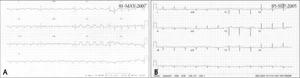
Fig. 1 A: Initial electrocardiogram showed low voltage, Q waves in inferior leads and ST-segment elevation in the leads V1 to V3. B: Previous electrocardiogram checked twenty months ago revealed no ST-segment elevation in the V1 to V3.

Fig. 2 A: The echocardiogram revealed external mass (white arrow) at the anterior side of the right ventricle. The mass invaded to the right ventricle and apical septum. B: The computerized tomography demonstrated external mass (black arrow) of the left chest wall and the mass invade to the right ventricle and apical portion of the interventricular septum.
Reference
-
1. Wang k, Asinger RW, Marriott HJ. ST-segment elevation in conditions other than acute myocardial infarction. N Engl J Med. 2007. 356:47–54.
Article
- Full Text Links
-
- Actions
-
Cited
- CITED
-
- Close
- Share
-
- Similar articles
-
- ST segment
- A Case of ST-Segment Elevation in a Patient with Subarachnoid Hemorrhage
- Acute Myocardial Infarction by Right Coronary Artery Occlusion Presenting as Precordial ST Elevation on Electrocardiography
- Erratum: Etiologies and Predictors of False-Positive Diagnosis of ST-Segment Elevation Myocardial Infarction
- ST Segment Elevation in Lead V1on Treadmill Exercise Test in the Patients with Angina : A Predictor of Coronary Artery Disease and It's Location
