Cochlear Implantation after Bilateral Transverse Temporal Bone Fractures
- Affiliations
-
- 1Department of Otolaryngology, Daegu Fatima Hospital, Daegu, Korea. sungheekim@fatima.or.kr
- KMID: 1486090
- DOI: http://doi.org/10.3342/ceo.2008.1.3.171
Abstract
- Patients deafened by a severe head injury are rarely encountered. We report a case of a 65-yr-old man with bilateral transverse temporal bone fractures due to head injury. He underwent cochlear implant and achieved a satisfactory auditory rehabilitation. Imaging studies of temporal bone before performing a cochlear implantation provide important information on a patient with bilateral temporal bone fractures. Cochlear implantations with careful planning in such a patient may be a very effective method for aural rehabilitation.
MeSH Terms
Figure
-

Fig. 1 (A) Axial CT scan of the temporal bones through the level of the otic capsules (bone window). Note the presence of bilateral transverse temporal bone fractures through the otic capsules (see black arrows). The temporal bone fractures appear to involve the otic capsule near the round window niches of the jugular bulb on the right side and through the cochlear axis on the left side. The left fracture line creates a bony separation shaped triangular particle posterior to the cochlear aqueduct and anterior to the jugular bulb and extends to the medial end of the internal auditory canal. Air can be seen in the middle turn of the cochlea, indicating a pneumolabyrinth (white arrow). (B) Axial CT scan through the level of the lateral semicircular canal. The fracture line on the right side involves the ampulla of the lateral semicircular canal and connects to the orifice of the vestibular aqueduct. Air can be seen in the right utricle and in the left internal auditory canal. Also there are effusions in the well pneumatized mastoid cavities, bilaterally, and in the left sphenoid sinus.
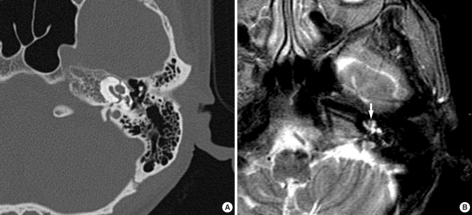
Fig. 2 Follow up imaging studies three months after the injury. (A) An enlarged axial CT scan of the left temporal bone through the level of the otic capsule shows no visible bony obstruction in the cochlear duct. (B) An enlarged axial T2 weighted MR scan shows a clearly visible fracture line with a hyposignal intensity which suggests fibrosis (white arrow).
Reference
-
1. Morgan WE, Coker NJ, Jenkins HA. Histopathology of temporal bone fractures: implications for cochlear implantation. Laryngoscope. 1994; 4. 104(4):426–432. PMID: 8164481.2. Camilleri AE, Toner JG, Howarth KL, Hampton S, Ramsden RT. Cochlear implantation following temporal bone fracture. J Laryngol Otol. 1999; 5. 113(5):454–457. PMID: 10505161.
Article3. Simons JP, Whitaker ME, Hirsch BE. Cochlear implantation in a patient with bilateral temporal bone fractures. Otolaryngol Head Neck Surg. 2005; 5. 132(5):809–811. PMID: 15886642.
Article4. Nadol JB Jr, Young YS, Glynn RJ. Survival of spiral ganglion cells in profound sensorineural hearing loss: implications for cochlear implantation. Ann Otol Rhinol Laryngol. 1989; 6. 98(6):411–416. PMID: 2729822.
Article5. Fredrickson JM, Griffith AW, Lindsay JR. Transverse fracture of the temporal bone. A clinical and histopathologic study. Arch Otolaryngol. 1963; 12. 78:770–784. PMID: 14059364.
- Full Text Links
-
- Actions
-
Cited
- CITED
-
- Close
- Share
-
- Similar articles
-
- Cochlear Implantation in a Deaf Patient with Bilateral Temporal Bone Fractures
- A Case of Cochlear Implantation after Bilateral Temporal Bone Fracture
- A Case of Cochlear Implantation Targeting Preserved Cerebral Cortex in Severe Traumatic Brain Injury
- A Case of Cochlear Implantation in a Postligual Deaf Patient with Osteogenesis Imperfecta
- Cochlear Implantation in the Cochlear Nerve Hypoplasia or Aplasia as Suggested by Temporal Bone Magnetic Resonance Imaging
