Pathological characterization of TgElk mice injected with brain homogenate from elk with chronic wasting disease
- Affiliations
-
- 1Ilsong Institute of Life Science, Hallym University, Anyang 431-060, Korea. yskim@hallym.ac.kr
- 2Department of Microbiology, College of Medicine, Hallym University, Chuncheon 200-702, Korea.
- 3New York State Institute for Basic Research in Developmental Disabilities, NY 10314, USA.
- KMID: 1482809
- DOI: http://doi.org/10.4142/jvs.2013.14.1.21
Abstract
- Chronic wasting disease (CWD) is classified as a transmissible spongiform encephalopathy or prion disease that affects cervids. CWD has been reported in 15 US states, two Canadian provinces, and in imported elk on several farms in Korea. This study was conducted to examine the molecular biological and pathogenic characteristics of a CWD-associated prion isolated in Korea. The epidemiological origin of this pathogen was also determined. Homozygous TgElk mice were infected with a CWD-affected elk brain pool prepared from the brain of an imported Canadian elk. We measured the incubation time of the pathogen, neuropathological changes by immunohistochemical staining, the pattern(s) of scrapie prion protein (PrPSc) deposition, and PrPSc protein profiles by Western blotting. We found that TgElk mice infected with brain homogenate from the elk suffering from CWD showed incubation times, vacuolar degeneration, and PrPSc accumulation similar to those previously reported in the literature. Our results suggest that homozygous TgElk mice efficiently transmit CWD with short incubation times and that this animal can serve a valuable research model and reliable in vivo diagnostic tool.
Keyword
MeSH Terms
Figure
-
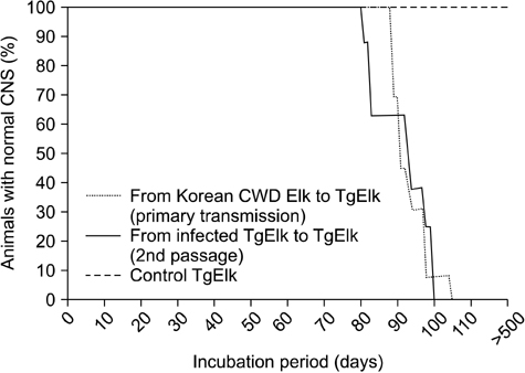
Fig. 1 Survival curves of cervidized Tg mice (TgElk) mice after injection of brain homogenates from chronic wasting disease (CWD)-positive elk and from CWD-positive TgElk mice (second passage).

Fig. 2 Pathological findings in brains of TgElk mice infected with brain homogenate from CWD-positive elk. Sections from the brain at the level of the hippocampus of an age-matched healthy control (A~E) and a Korean CWD isolate-infected TgElk mouse (F~O). Each section was stained with hematoxylin and eosin or subjected to immunohistochemistry to monitor the presence glial fibrillary acidic protein, CD11b, and PrP. CC: corpus callosum, CA3: cornu ammonis3, arrows: vacuole, asterisks: florid plaque. Scale bars = 500 µm (B, C, D, E, G, H, I, J, M, and N), and 50 µm (K, L, and O).

Fig. 3 Scrapie prion protein (PrPSc) Western blot patterns of brain homogenate from CWD-positive elk and infected TgElk mice. Lanes 1 and 2: homogenates from the donor elk with CWD; lanes 3~8: three control TgElk mice brains; lanes 9~16: four infected TgElk mice brains. '-': untreated brain homogenates, '+': brain homogenates treated with proteinase K (PK).

Fig. 4 Comparison of the vacuolar lesion profiles for TgElk mice injected with brain homogenate from CWD-positive elk from Korea or mule deer from the USA.
Reference
-
1. Browning SR, Mason GL, Seward T, Green M, Eliason GAJ, Mathiason C, Miller MW, Williams ES, Hoover E, Telling GC. Transmission of prions from mule deer and elk with chronic wasting disease to transgenic mice expressing cervid PrP. J Virol. 2004. 78:13345–13350.
Article2. Bruce ME, Fraser H, McBride PA, Scott JR, Dickinson AG. Prusiner SB, Collinge J, Powell J, Anderton B, editors. The basis of strain variation in scrapie. Prion Diseases of Humans and Animals. 1992. New York: Ellis Horwood;497–508.3. Choi JK, Park SJ, Jun YC, Oh JM, Jeong BH, Lee HP, Park SN, Carp RI, Kim YS. Generation of monoclonal antibody recognized by the GXXXG motif (glycine zipper) of prion protein. Hybridoma (Larchmt). 2006. 25:271–277.
Article4. Kim TY, Shon HJ, Joo YS, Mun UK, Kang KS, Lee YS. Additional cases of chronic wasting disease in imported deer in Korea. J Vet Med Sci. 2005. 67:753–759.
Article5. Kim YS, Carp RI, Callahan SM, Natelli M, Wisniewski HM. Vacuolization, incubation period and survival time analyses in three mouse genotypes injected stereotactically in three brain regions with the 22L scrapie strain. J Neuropathol Exp Neurol. 1990. 49:106–113.
Article6. Kong Q, Huang S, Zou W, Vanegas D, Wang M, Wu D, Yuan J, Zheng M, Bai H, Deng H, Chen K, Jenny AL, O'Rourke K, Belay ED, Schonberger LB, Petersen RB, Sy MS, Chen SG, Gambetti P. Chronic wasting disease of elk: transmissibility to humans examined by transgenic mouse models. J Neurosci. 2005. 25:7944–7949.
Article7. LaFauci G, Carp RI, Meeker HC, Ye X, Kim JI, Natelli M, Cedeno M, Petersen RB, Kascsak R, Rubenstein R. Passage of chronic wasting disease prion into transgenic mice expressing Rocky Mountain elk (Cervus elaphus nelsoni) PrPC. J Gen Virol. 2006. 87(Pt 12):3773–3780.
Article8. McLean AR, Bostock CJ. Scrapie infections initiated at varying doses: an analysis of 117 titration experiments. Philos Trans R Soc Lond B Biol Sci. 2000. 355:1043–1050.
Article9. Miller MW, Williams ES. Chronic wasting disease of cervids. Curr Top Microbiol Immunol. 2004. 284:193–214.
Article10. Raymond GJ, Raymond LD, Meade-White KD, Hughson AG, Favara C, Gardner D, Williams ES, Miller MW, Race RE, Caughey B. Transmission and adaptation of chronic wasting disease to hamsters and transgenic mice: evidence for strains. J Virol. 2007. 81:4305–4314.
Article11. Sohn HJ, Kim JH, Choi KS, Nah JJ, Joo YS, Jean YH, Ahn SW, Kim OK, Kim DY, Balachandran A. A case of chronic wasting disease in an elk imported to Korea from Canada. J Vet Med Sci. 2002. 64:855–858.
Article12. Tamgüney G, Giles K, Bouzamondo-Bernstein E, Bosque PJ, Miller MW, Safar J, DeArmond SJ, Prusiner SB. Transmission of elk and deer prions to transgenic mice. J Virol. 2006. 80:9104–9114.
Article
- Full Text Links
-
- Actions
-
Cited
- CITED
-
- Close
- Share
-
- Similar articles
-
- Development of monoclonal antibodies against the abnormal prion protein isoform (PrPres) associated with chronic wasting disease (CWD)
- Elk dander-induced occupational asthma
- HPV-18 E7 Interacts with Elk-1 Leading to Elevation of the Transcriptional Activity of Elk-1 in Cervical Cancer
- A Case of Cerebral salt Wasting Syndrome with Pseudomonas Meningitis after Removal of Pituitary Adenoma
- Cerebral salt wasting syndrome caused by external lumbar drainage in a patient with chronic hydrocephalus
