CT-Guided Core Needle Biopsy of Deep Suprahyoid Head and Neck Lesions
- Affiliations
-
- 1Department of Medical Imaging and Internvetion, Chang Gung Memorial Hospital, College of Medicine, Chang Gung University, Taoyuan 333, Taiwan. shng@adm.cgmh.org.tw
- KMID: 1482791
- DOI: http://doi.org/10.3348/kjr.2013.14.2.299
Abstract
OBJECTIVE
To evaluate the efficacy of computer tomography (CT)-guided core needle biopsy (CNB) in the diagnosis of deep suprahyoid lesions in patients with treated head and neck cancers.
MATERIALS AND METHODS
Between December, 2003 and May, 2011, 28 CT-guided CNBs were performed in 28 patients with deep suprahyoid head and neck lesions. All patients had undergone treatment for head and neck cancers. Subzygomatic, paramaxillary, and retromandibular approaches were used. The surgical results, response to treatment, and clinical follow-up were used as the diagnostic reference standards.
RESULTS
All biopsies yielded adequate specimens for definitive histological diagnoses. A specimen from a right parapharyngeal lesion showed atypia, which was deemed a false negative diagnosis. Diagnostic accuracy was 27/28 (96.4%). Two minor complications were encountered: a local hematoma and transient facial palsy. Between the 18 or 20 gauge biopsy needles, there was no statistical difference in the diagnostic results.
CONCLUSION
CT-guided core needle biopsy, with infrequent and minor complications, is an accurate and efficient method for the histological diagnosis of deep suprahyoid lesions in post-treated head and neck cancer patients. This procedure can preclude an unnecessary surgical intervention, especially in patients with head and neck cancers.
MeSH Terms
Figure
-
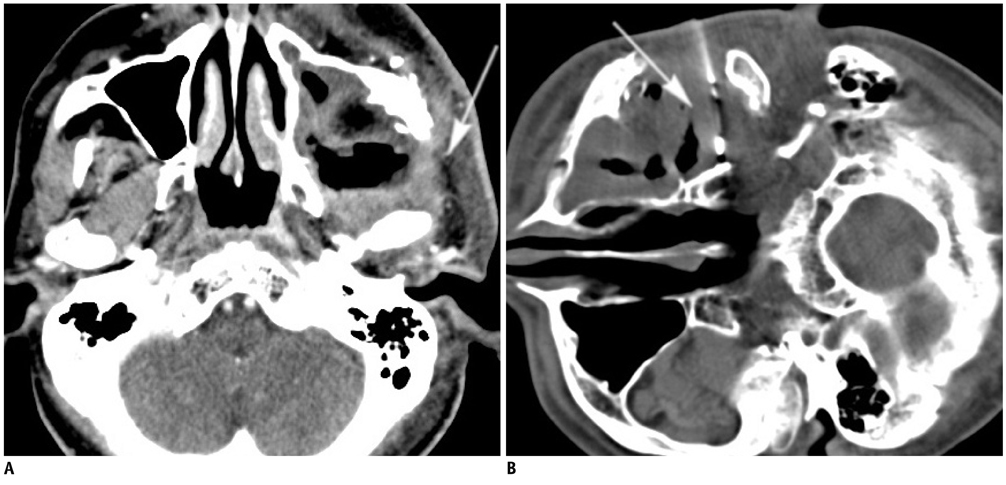
Fig. 1 Fifty one-year-old male (case 28) with left buccal cancer after surgery and radiotherapy. A. Contrast-enhanced CT scan shows large necrotic lesion in left masticator space with infiltrative part at periphery (arrow). B. Using subzygomatic approach, 17/18 G biopsy needle set is inserted into lesion (arrow). Biopsy revealed fibrosis with granulation. Follow-up image studies in three and six months showed lesion in stationary status, consistent with biopsy result (not shown).
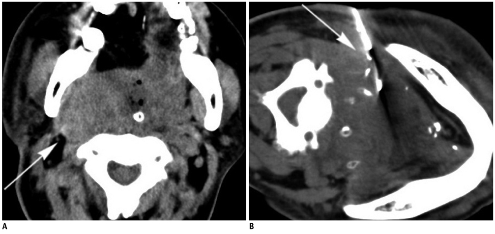
Fig. 2 Sixty four-year-old male (case 27) with history of tongue cancer after operation and radiotherapy. A. Pre-procedural CT scan shows large infiltrative lesion in right parapharyngeal space (arrow). B. Using retromandibular approach, 17/18 G biopsy needle set is inserted in lesion (arrow). Biopsy revealed atypia. Progressive enlargement of lesion was noted in follow-up CT in one month (not shown). Patient died two months after procedure due to poor condition.
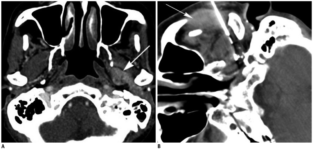
Fig. 3 Sixty four-year-old male (case 20) with buccal cancer after operation and radiotherapy. A. Contrast-enhanced CT shows infiltrative lesion in left parapharyngeal space (arrow). B. Using subzygomatic approach, 17/18 G biopsy needle set is inserted in lesion. Note hematoma in left messeter muscle (arrow). Biopsy revealed metastatic squamous cell carcinoma.

Fig. 4 Forty-year-old male (case 11) with left parotid adenoid cystic carcinoma after surgical excision. A. Contrast-enhanced CT shows heterogeneous hypodense lesion in deep lobe of left parotid gland (arrow). B. Using retromandibular approach, 17/18 G needle set is inserted in lesion (arrow). Biopsy revealed adenoid cystic carcinoma. Transient left facial palsy was noted immediately after procedure. Symptom subsided in 30 minutes.
Reference
-
1. Connor SE, Chaudhary N. CT-guided percutaneous core biopsy of deep face and skull-base lesions. Clin Radiol. 2008. 63:986–994.2. Nyquist GG, Tom WD, Mui S. Automatic core needle biopsy: a diagnostic option for head and neck masses. Arch Otolaryngol Head Neck Surg. 2008. 134:184–189.3. Gupta S, Henningsen JA, Wallace MJ, Madoff DC, Morello FA Jr, Ahrar K, et al. Percutaneous biopsy of head and neck lesions with CT guidance: various approaches and relevant anatomic and technical considerations. Radiographics. 2007. 27:371–390.4. DelGaudio JM, Dillard DG, Albritton FD, Hudgins P, Wallace VC, Lewis MM. Computed tomography--guided needle biopsy of head and neck lesions. Arch Otolaryngol Head Neck Surg. 2000. 126:366–370.5. Sherman PM, Yousem DM, Loevner LA. CT-guided aspirations in the head and neck: assessment of the first 216 cases. AJNR Am J Neuroradiol. 2004. 25:1603–1607.6. Howlett DC, Harper B, Quante M, Berresford A, Morley M, Grant J, et al. Diagnostic adequacy and accuracy of fine needle aspiration cytology in neck lump assessment: results from a regional cancer network over a one year period. J Laryngol Otol. 2007. 121:571–579.7. Pfeiffer J, Kayser G, Technau-Ihling K, Boedeker CC, Ridder GJ. Ultrasound-guided core-needle biopsy in the diagnosis of head and neck masses: indications, technique, and results. Head Neck. 2007. 29:1033–1040.8. Screaton NJ, Berman LH, Grant JW. Head and neck lymphadenopathy: evaluation with US-guided cutting-needle biopsy. Radiology. 2002. 224:75–81.9. Abrahams JJ. Mandibular sigmoid notch: a window for CT-guided biopsies of lesions in the peripharyngeal and skull base regions. Radiology. 1998. 208:695–699.10. Mukherji SK, Turetsky D, Tart RP, Mancuso AA. A technique for core biopsies of head and neck masses. AJNR Am J Neuroradiol. 1994. 15:518–520.11. Tu AS, Geyer CA, Mancall AC, Baker RA. The buccal space: a doorway for percutaneous CT-guided biopsy of the parapharyngeal region. AJNR Am J Neuroradiol. 1998. 19:728–731.12. Cheung JY, Kim Y, Shim SS, Lim SM. Combined fluoroscopy- and CT-guided transthoracic needle biopsy using a C-arm cone-beam CT system: comparison with fluoroscopy-guided biopsy. Korean J Radiol. 2011. 12:89–96.13. Tomozawa Y, Inaba Y, Yamaura H, Sato Y, Kato M, Kanamoto T, et al. Clinical value of CT-guided needle biopsy for retroperitoneal lesions. Korean J Radiol. 2011. 12:351–357.14. Renshaw AA, Pinnar N. Comparison of thyroid fine-needle aspiration and core needle biopsy. Am J Clin Pathol. 2007. 128:370–374.15. Charboneau JW, Reading CC, Welch TJ. CT and sonographically guided needle biopsy: current techniques and new innovations. AJR Am J Roentgenol. 1990. 154:1–10.16. Walker AT, Chaloupka JC, Putman CM, Abrahams JJ, Ross DA. Sentinel transoral hemorrhage from a pseudoaneurysm of the internal maxillary artery: a complication of CT-guided biopsy of the masticator space. AJNR Am J Neuroradiol. 1996. 17:377–381.
- Full Text Links
-
- Actions
-
Cited
- CITED
-
- Close
- Share
-
- Similar articles
-
- Ultrasound of Head and Neck: Anatomy
- Diagnostic Utility of Ultrasound-Guided Core-Needle Biopsy Performed by a Head and Neck Surgeon for Mass Lesions with Inconclusive Result in Fine-Needle Aspiration Cytology
- Breast Lesions with Discordant Results on Ultrasound-guided Core Needle Biopsy
- Usefulness of Ultrasound-Guided Automated Core Biopsy of Nonpalpable Breast Lesions
- Two Cases of Thyroid Hematoma Developing after a Core Needle Biopsy
