Insertion and removal torques according to orthodontic mini-screw design
- Affiliations
-
- 1Department of Orthodontics, College of Dentistry, Yonsei University, Korea.
- 2Department of Orthodontics, College of Dentistry, Dental Science Research Institute, Yonsei University, Korea. hwang@yuhs.ac
- KMID: 1481501
- DOI: http://doi.org/10.4041/kjod.2008.38.1.5
Abstract
OBJECTIVE
This study was designed to analyze the primary and secondary stability characteristics of orthodontic mini-screws of tapered design when compared with the cylinder mini-screw.
METHODS
A total of 48 mini-screws were placed into the buccal alveolar bone of the mandible in 6 male beagle dogs. Comparison was made between tapered and cylinder type mini-screws (Biomaterials Korea, Seoul, Korea). Maximum insertion torque (MIT) was measured using a torque sensor (Mark-10, MGT 50, USA) during installation, and maximum removal torque (MRT) was recorded after 3 and 12 weeks of loading.
RESULTS
Taper mini-screws showed a higher MIT value of 22.3 Ncm compared with cylinder mini-screw showing 13.6 Ncm (p < 0.001). The MRT of the taper mini-screw showed a significantly higher value of 9.1 Ncm than those of cylinder mini-screw of 5.7 Ncm at 3-weeks after installation (p < 0.05). However, there was no difference in the MRT value between the taper and cylinder mini-screws at 12 weeks of loading.
CONCLUSIONS
These results showed that the high insertion torque of the taper mini-screw design increases initial stability until 3 weeks of loading, but does not have any effect on the secondary stability at 12 weeks of loading.
Figure
-
Fig 1 Drawing of cylindrical type (1507C) & taper type (1507T) mini-screws (unit: mm).
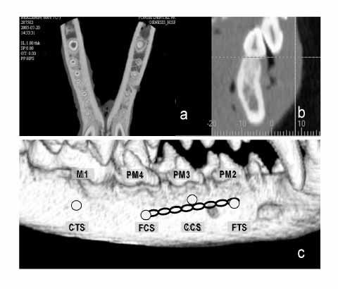
Fig 2 Schematic image for mini-screw insertion. a and b, Axial and sagittal images for the localization of mini-screws; c, force applied groups were reciprocally loaded by elastic-chain. FCS, force applied cylinder mini-screw; FTS, force applied taper mini-screw; CCS, control group of cylinder mini-screw; CTS, control group of taper mini-screw.
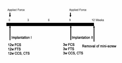
Fig 3 Timetable for placing mini-screw. w, weeks; FCS, force applied cylindrical mini-screw; FTS, force applied taper mini-screw; CCS, control group of cylindrical mini-screw; CTS, control group of taper mini-screw.
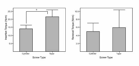
Fig 4 Insertion and removal torque (Ncm) for screw types. Statistically significant difference between periods by independent t-test; *p < 0.001.

Fig 5 Graphs of insertion and removal torque for loading periods. Statistically significant difference between periods by independent t-test and Scheffe test, *p < 0.05; †p < 0.01; ‡p < 0.001.
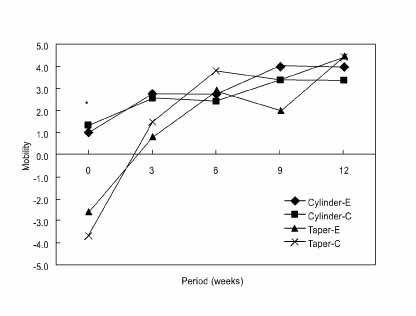
Fig 6 Graph of mobility change for loading periods. Cylinder-E, Cylinder type of experimental group; Cylinder-C, cylinder type of control group; Taper-E, taper type of experimental group; Taper-C, taper type of control group, *p < 0.05.
Cited by 4 articles
-
Effects of surface treatment on the osseointegration potential of orthodontic mini-implant
Mi-sun Jeon, Yoon-Goo Kang, Sung-Seo Mo, Keun-Hye Lee, Yoon-Ah Kook, Seong-Hun Kim
Korean J Orthod. 2008;38(5):328-336. doi: 10.4041/kjod.2008.38.5.328.Finite element analysis of cortical bone strain induced by self-drilling placement of orthodontic microimplant
Jin-Seo Park, Wonjae Yu, Hee-Moon Kyung, Oh-Won Kwon
Korean J Orthod. 2009;39(4):203-212. doi: 10.4041/kjod.2009.39.4.203.The effects of different pilot-drilling methods on the mechanical stability of a mini-implant system at placement and removal: a preliminary study
Il-Sik Cho, HyeRan Choo, Seong-Kyun Kim, Yun-Seob Shin, Duck-Su Kim, Seong-Hun Kim, Kyu-Rhim Chung, John C. Huang
Korean J Orthod. 2011;41(5):354-360. doi: 10.4041/kjod.2011.41.5.354.Cortical bone strain during the placement of orthodontic microimplant studied by 3D finite element analysis
Okhyun Nam, Wonjae Yu, Hee-Moon Kyung
Korean J Orthod. 2008;38(4):228-298. doi: 10.4041/kjod.2008.38.4.238.
Reference
-
1. Chen CH, Chang CS, Hsieh CH, Tseng YC, Shen YS, Huang IY, et al. The use of microimplants in orthodontic anchorage. J Oral Maxillofac Surg. 2006. 64:1209–1213.
Article2. Buchter A, Wiechmann D, Koerdt S, Wiesmann HP, Piffko J, Meyer U. Load-related implant reaction of mini-implants used for orthodontic anchorage. Clin Oral Implants Res. 2005. 16:473–479.
Article3. Joos U, Buchter A, Wiesmann HP, Meyer U. Strain driven fast osseointegration of implants. Head Face Med. 2005. 1:6.
Article4. Huja SS, Litsky AS, Beck FM, Johnson KA, Larsen PE. Pull-out strength of monocortical screws placed in the maxilla and mandibles of dogs. Am J Orthod Dentofacial Orthop. 2005. 127:307–313.
Article5. Brown GA, McCarthy T, Bourgeault CA, Callahan DJ. Mechanical performance of standard and cannulated 4.0-mm cancellous bone screws. J Orthop Res. 2000. 18:307–312.
Article6. Heidemann W, Gerlach KL, Grobel KH, Kollner HG. Influence of different pilot hole sizes on torque measurements and pullout analysis of osteosynthesis screws. J Craniomaxillofac Surg. 1998. 26:50–55.
Article7. Miyawaki S, Koyama I, Inoue M, Mishima K, Sugahara T, Takano-Yamamoto T. Factors associated with the stability of titanium screws placed in the posterior region for orthodontic anchorage. Am J Orthod Dentofacial Orthop. 2003. 124:373–378.
Article8. Cheng SJ, Tseng IY, Lee JJ, Kok SH. A prospective study of the risk factors associated with failure of mini-implants used for orthodontic anchorage. Int J Oral Maxillofac Implants. 2004. 19:100–106.9. Motoyoshi M, Hirabayashi M, Uemura M, Shimizu N. Recommended placement torque when tightening an orthodontic mini-implant. Clin Oral Implants Res. 2006. 17:109–114.
Article10. Meredith N. Assessment of implant stability as a prognostic determinant. Int J Prosthodont. 1998. 11:491–501.11. Ueda M, Matsuki M, Jacobsson M, Tjellstrom A. Relationship between insertion torque and removal torque analyzed in fresh temporal bone. Int J Oral Maxillofac Implants. 1991. 6:442–447.12. Frost HM. Bone's mechanostat: a 2003 update. Anat Rec A Discov Mol Cell Evol Biol. 2003. 275:1081–1101.
Article13. Frost HM. A brief review for orthopedic surgeons: fatigue damage (microdamage) in bone (its determinants and clinical implications). J Orthop Sci. 1998. 3:272–281.
Article14. Sowden D, Schmitz JP. AO self-drilling and self-tapping screws in rat calvarial bone: An ultrastructural study of the implant interface. J Oral Maxillofac Surg. 2002. 60:294–299.
Article15. Degidi M, Scarano A, Petrone G, Piattelli A. Histologic analysis of clinically retrieved immediately loaded titanium implants: a report of 11 cases. Clin Implant Dent Relat Res. 2003. 5:89–93.
Article16. Degidi M, Scarano A, Iezzi G, Piattelli A. Periimplant bone in immediately loaded titanium implants: histologic and histomorphometric evaluation in human. A report of two cases. Clin Implant Dent Relat Res. 2003. 5:170–175.
Article17. Melsen B, Lang NP. Biological reactions of alveolar bone to orthodontic loading of oral implants. Clin Oral Implants Res. 2001. 12:144–152.
Article18. Oh NH, Kim SH, Kook YA, Lee KH, Kang YG, Mo SS. Removal torque of sandblasted large grit and acid etched treated mini-implant. Korean J Orthod. 2006. 36:324–330.19. Branemark R, Ohrnell L-O, Nilsson P, Thomsen P. Biomechanical characterization of osseointegration during healing: an experimental in vivo study in the rat. Biomaterials. 1997. 18:969–978.
Article20. Lee JH, Ryu HS, Lee DS, Hong KS, Chang BS, Lee CK. Biomechanical and histomorphometric study on the bone-screw interface of bioactive ceramic-coated titanium screws. Biomaterials. 2005. 26:3249–3257.
Article21. Park HS, Yen S, Jeoung SH. Histologic and biomechanical characteristics of orthodontic self-drilling and self-tapping microscrew implants. Korean J Orthod. 2006. 36:16–29.22. Chen YJ, Chen YH, Lin LD, Yao CC. Removal torque of miniscrews used for orthodontic anchorage--a preliminary report. Int J Oral Maxillofac Implants. 2006. 21:283–289.23. Simon H, Caputo AA. Removal torque of immediately loaded transitional endosseous implants in human subjects. Int J Oral Maxillofac Implants. 2002. 17:839–845.24. Schulte W, Lukas D. The Periotest method. Int Dent J. 1992. 42:433–440.
- Full Text Links
-
- Actions
-
Cited
- CITED
-
- Close
- Share
-
- Similar articles
-
- Effect of dual pitch mini-implant design and diameter of an orthodontic mini-implant on the insertion and removal torque
- Geometrical design characteristics of orthodontic mini-implants predicting maximum insertion torque
- Comparison of success rates of orthodontic mini-screws by the insertion method
- Evaluation of insertion of torque and pull-out strength of mini-screws according to different thickness of artificial cortical bone
- Effect of cutting flute length and shape on insertion and removal torque of orthodontic mini-implants
