Labial and buccal surface contours of Korean normal occlusion in a three-dimensional digital model
- Affiliations
-
- 1Department of Orthodontics, School of Dentistry, Yonsei University, Korea.
- 2HT Corp., Korea.
- 3Department of Orthodontics, School of Dentistry, Dental Science Research Institute, Yonsei University, Korea. ypark@yuhs.ac
- KMID: 1481497
- DOI: http://doi.org/10.4041/kjod.2008.38.2.95
Abstract
- OBJECTVIE: This study was performed to investigate the labio/buccal clinical crown curvatures of Korean permanent teeth and to obtain the curve-ratio data in an attempt to fabricate bracket bases fit for each individual Korean permanent tooth.
METHODS
Three-dimensional digital models were made from 30 sets of dental casts with normal anatomic structures. According to the FA points, horizontal and vertical reference planes were established and lines were drawn on the tooth surfaces in reference to these planes. The curvature was expressed as the coefficient of a quadratic equation. Lines mesial, distal, gingival and occlusal to the horizontal, vertical reference planes and the FA point were drawn.
RESULTS
The curvature measured for each line revealed that there are no significant differences between male and female, except for maxillary canines and maxillary second bicuspids (p > 0.05). There were notable differences in the mesio-distal or gingivo-occlusal curvatures among the mandibular lateral incisors, maxillary canines, maxillary and mandibular first and second bicuspids and first molars (p < 0.05).
CONCLUSIONS
The labial & buccal crown curvatures of teeth in Korean normal occlusion were measured on the mesial and distal, gingival and occlusal sides respectively in this study. Based on these data, a SWA can be developed to fit the individual features of Korean tooth crowns.
Keyword
Figure
-
Fig 1 Orapix KOD-300 scanner.
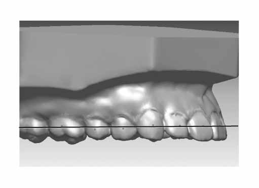
Fig 2 The establishment of horizontal reference plane at maxilla. Some FA points may not be on the horizontal reference plane due to average calculation.

Fig 3 The establishment of horizontal and vertical reference plane at maxillary first premolar.
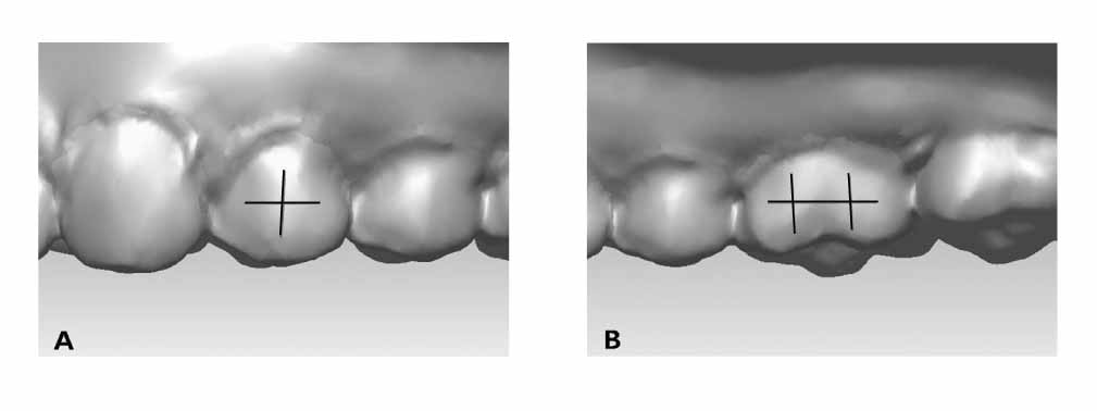
Fig 4 The establishment of horizontal and vertical reference curves at maxillary first premolar (A) and first molar (B).
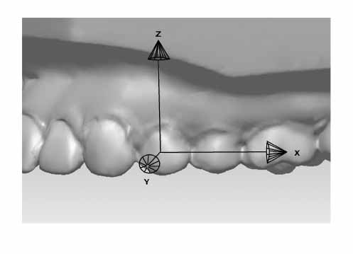
Fig 5 The features which forms axis of coordinates on maxillary second premolar. X and Y axes on the horizontal reference plane have the same Z value which is 0.
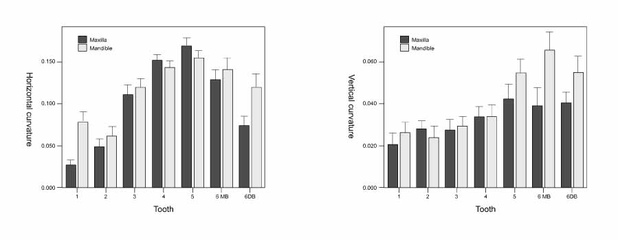
Fig 6 Graphs of crown curvatures in each reference plane. Left, Labial/buccal curvatures; Right, occluso/gingival curvatures. Bar show means, and error bars show 95.0% confidence limit of mean. MB, Mesiobuccal surface; DB, distobuccal surface.
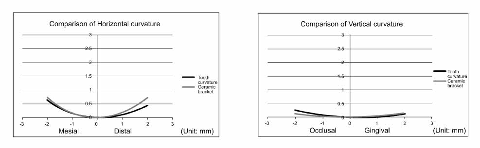
Fig 7 Comparison of the ceramic bracket (Miso, HT, Ansan, Korea) curvatures and the mean value curvatures in horizontal (left) and vertical (right) plane at maxillary second premolar.
Cited by 2 articles
-
Comparison of model analysis measurements among plaster model, laser scan digital model, and cone beam CT image
Mi-young Lim, Sung-hoon Lim
Korean J Orthod. 2009;39(1):6-17. doi: 10.4041/kjod.2009.39.1.6.New classification of lingual arch form in normal occlusion using three dimensional virtual models
Kyung Hee Park, Mohamed Bayome, Jae Hyun Park, Jeong Woo Lee, Seung-Hak Baek, Yoon-Ah Kook
Korean J Orthod. 2015;45(2):74-81. doi: 10.4041/kjod.2015.45.2.74.
Reference
-
1. Graber TM, Vanarsdall RL. Orthodontics: Current principles and techniques. 1994. St Louis: Mosbys;659–663.2. Andrews LF. The six keys to normal occlusion. Am J Orthod. 1972. 62:296–309.
Article3. Andrews LF. The straight-wire appliance, origin, controversy, commentary. J Clin Orthod. 1976. 10:99–114.4. Andrews LF. The straight-wire appliance. Explained and compared. J Clin Orthod. 1976. 10:174–195.5. Roth RH. Five year clinical evaluation of the Andrews straight-wire appliance. J Clin Orthod. 1976. 10:836–850.6. Roth RH. The straight-wire appliance 17 years later. J Clin Orthod. 1987. 21:632–642.7. Roth RH. Functional occlusion for the Orthodontist. Part III. J Clin Orthod. 1981. 15:174–179. 182–198.8. McLaughlin RP, Bennett JC. Bracket placement with the preadjusted appliance. J Clin Orthod. 1995. 29:302–311.9. Sperry TP, Worms FW, Isaacson RJ, Speidel TM. Tooth-size discrepancy in mandibular prognathism. Am J Orthod. 1977. 72:183–190.
Article10. Nie Q, Lin J. Comparison of intermaxillary tooth size discrepancies among different malocclusion groups. Am J Orthod Dentofacial Orthop. 1999. 116:539–544.
Article11. Kim DS, Kim YJ, Choi JH, Han JH. A Study of Korean Norm about tooth size and ratio in Korean adults with normal occlusion. Korean J Orthod. 2001. 31:505–515.12. Lee SJ, Moon SC, Kim TW, Nahm DS, Chang YI. Tooth size and arch parameters of normal occlusion in a large Korean sample. Korean J Orthod. 2004. 34:473–480.13. Nojima K, McLaughlin RP, Isshiki Y, Sinclair PM. A comparative study of Caucasian and Japanese mandibular clinical arch forms. Angle Orthod. 2001. 71:195–200.14. Kook YA, Nojima K, Moon HB, McLaughlin RP, Sinclair PM. Comparison of arch forms between Korean and North American white populations. Am J Orthod Dentofacial Orthop. 2004. 126:680–686.
Article15. Germane N, Bentley BE Jr, Isaacson RJ. Three biologic variables modifying faciolingual tooth angulation by straight-wire appliances. Am J Orthod Dentofacial Orthop. 1989. 96:312–319.
Article16. Vigorito JW, Moresca R, Dominguez GC, Tortamano A. Influence of the convexity of the upper central incisor on the torque expression of preadjusted brackets. J Clin Orthod. 2006. 40:42–46.17. van Loenen M, Degrieck J, De Pauw G, Dermaut L. Anterior tooth morphology and its effect on torque. Eur J Orthod. 2005. 27:258–262.
Article18. Pak YC. A morphologic study on straight wire bracket for Korean. Korean J Orthod. 1991. 21:481–493.19. Yoon JJ, Sohn BH. A study of the crown angulation in normal occlusion. Korean J Orthod. 1986. 14:123–165.20. Chang YI, Yang YS, Nahm DS, Moon SC. Effects A Study for the development of the Korean orthodontic bracket. Korean J Orthod. 2000. 30:565–578.21. Kim SC, Nahm DS. A study on the configurations of Korean normal dental arches for preformed archwire. Korean J Orthod. 1984. 14:93–101.22. Lee DJ. Oriental Bracket. Korean J Orthod. 1991. 21:495–500.23. Kim JS, Jin KH, Hong SJ. A statistical study of clinical crown inclination in Korean's naturally occurring optimal occlusion. Korean J Orthod. 1992. 22:715–732.24. Lee WY, Park YC, Lim KS. A Morphometric study of teeth on the Korean normal occlusion. Korean J Orthod. 1998. 28:601–609.25. Kuroda T, Motohashi N, Tominaga R, Iwata K. Three-dimensional dental cast analyzing system using laser scanning. Am J Orthod Dentofacial Orthop. 1996. 110:365–369.
Article26. Quimby ML, Vig KW, Rashid RG, Firestone AR. The accuracy and reliability of measurements made on computer-based digital models. Angle Orthod. 2004. 74:298–303.27. Miethke RR, Melsen B. Effect of variation in tooth morphology and bracket position on first and third order correction with preadjusted appliances. Am J Orthod Dentofacial Orthop. 1999. 116:329–335.
Article28. Ko SD, Cha KS. A Study on the labial & buccal surface contour in Korean permanent teeth using three-dimensional laser scanning. Korean J Orthod. 2002. 32:275–291.29. Kim HJ. Dental morphology. 2007. Seoul: Hyunmoon;95–122.
- Full Text Links
-
- Actions
-
Cited
- CITED
-
- Close
- Share
-
- Similar articles
-
- The effect of labial inclination on intrusion of the upper and lower incisors by three-dimensional finite element analysis
- Three dimensional photoelastic study on the initial stress distributions of alveolar bone when retracted by lingual K-loop archwire
- A Study on the labial & buccal surface contour in Korean permanent teeth using three-dimensional laser scanning
- Three Dimensional Automatic Surface Reconstruction Software
- Differences in molar relationships and occlusal contact areas evaluated from the buccal and lingual aspects using 3-dimensional digital models
