Catamenial Hemoptysis Treated by Video-assisted Thoracoscopic Surgery
- Affiliations
-
- 1Department of Internal Medicine, Hallym University College of Medicine, Chuncheon, Korea. dongyu@hallym.ac.kr
- 2Department of Pathology, Hallym University College of Medicine, Chuncheon, Korea.
- 3Department of Thoracic and Cardiovascular Surgery, Hallym University College of Medicine, Chuncheon, Korea.
- KMID: 1478213
- DOI: http://doi.org/10.4046/trd.2008.65.1.29
Abstract
- Catamenial hemoptysis is a rare condition that's characterized by recurrent hemoptysis occurring in association with menstruation, and this is associated with the presence of intrapulmonary or endobronchial endometrial tissue. The diagnosis of pulmonary endometriosis can be made according to a typical clinical history and with exclusion of other causes of recurrent hemoptysis. Treatment of pulmonary endometriosis can be medical or surgical; however, the optimal management of this condition is still a matter of debate. Medical therapy may be problematic, due to recurrence of symptoms despite hormonal ablation, and adverse effects from long-term hormone therapy can also be a problem. We report here on a case of pulmonary endometriosis in a 23-year-old woman who presented with hemoptysis that occurred during the first 3 days of menstruation, and this happened over a 4 month period. She was successfully treated by video-assisted thoracoscopic surgery (VATS). No more hemoptysis was noted during 12 months of follow-up.
MeSH Terms
Figure
-

Figure 1 Preoperative chest x-ray shows no abnormal finding.
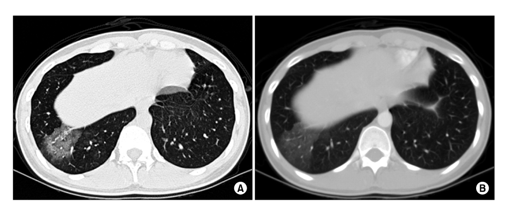
Figure 2 A chest HRCT image obtained during the patient's menstrual period shows consolidation and ground glass opacity at the lateral and posterior basal segment of right lower lobe (A). 2 months later, the other chest CT image shows aggravation of consolidation and ground glass opacity at the same segments (B).

Figure 3 Engorged blood vessels were noted in the right lower lobar bronchus (A). 2 months later, a small amount of fresh blood was coming out from the basal segment of the right lower lobe (B, C).
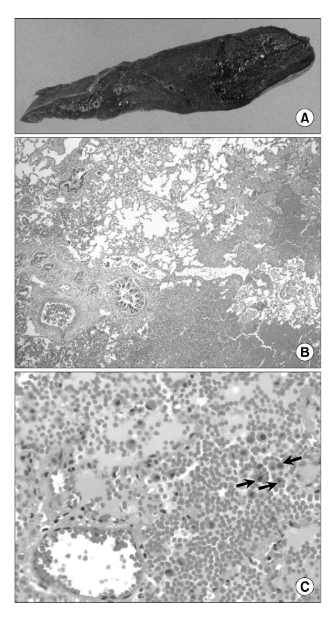
Figure 4 A lobectomy specimen of right lower lung, measuring 15×10×5 cm, reveals a pooly-demarcated hemorrhagic mass (4×3.5×1.5 cm), 10 cm apart from the bronchial resection margin (A). Microscopically, there is extensive intraalveolar hemorrhage with congested dilated vessels (H&E stain, ×40) (B). In the hemorrhagic alveolar spaces, many hemosiderin-laden macrophages (arrows) are seen (H&E stain, ×400) (C).
Reference
-
1. Joseph J, Sahn SA. Thoracic endometriosis syndrome: new observations from an analysis of 110 cases. Am J Med. 1996. 100:164–170.2. Wood DJ, Krishnan K, Stocks P. Catamenial hemoptysis: a rare cause. Thorax. 1993. 48:1048–1049.3. Park WW. Experimental trophoblastic embolism of the lungs. J Pathol Bacteriol. 1958. 75:257–265.4. Chung SY, Kim SJ, Kim TH, Ryu WG, Park SJ, Lee DY, et al. Computed tomography findings of pathologically confirmed pulmonary parenchymal endometriosis. J Comput Assist Tomogr. 2005. 29:815–818.5. Cassina PC, Hauser M, Kacl G, Imthurn B, Schroder S, Weder W. Catamenial hemoptysis. Diagnosis with MRI. Chest. 1997. 111:1447–1450.6. Kuo PH, Wang HC, Liaw YS, Kuo SH. Bronchoscopic and angiographic findings in tracheobronchial endometriosis. Thorax. 1996. 51:1060–1061.7. Katoh O, Yamada H, Aoki Y, Matsumoto S, Kudo S. Utility of angiograms in patients with catamenial hemoptysis. Chest. 1990. 98:1296–1297.8. Kim JH, Joo YT, Ahn OJ, Jeon SW, Moon Y, Choi JY, et al. A case of catamenial hemoptysis. Korean J Obset Gynecol. 2005. 48:500–504.9. Kim DH, Suh YA, Kim SI, Choi KS, Son HB, Lee JC, et al. A Case of catamenial hemoptysis treated successfully with gonadotropin-releasing hormone (GnRH) analogue. Tuberc Respir Dis. 2002. 53:349–353.10. Ham SH, Chung MP, Lee BW, Han KH, Kim HJ, Kwon OJ, et al. A case of pulmonary endometriosis resected by video-assisted thoracoscopic surgery. Tuberc Respir Dis. 2004. 56:542–549.11. Cho SJ, Ryu SM, Kim WJ, Lee SJ, Kim YS. Video-assisted thoracic surgery for pulmonay endometriosis: report of 1 case. Tuberc Respir Dis. 2006. 60:576–580.12. Park SM, Shin EJ, Kang KM, Kim MK, Cho DG, Kim CH. A case of pulmonary endometriosis treated by resection. Tuberc Respir Dis. 2006. 61:394–397.13. Park YB, Heo GM, Moon HK, Cho SJ, Shin YC, Eom KS, et al. Pulmonary endometriosis resected by video-assisted thoracoscopic surgery. Respirology. 2006. 11:221–223.14. Puma F, Carloni A, Casucci G, Puligheddu C, Urbani M, Porcaro G. Successful endoscopic Nd-YAG laser treatment of endobronchial endometriosis. Chest. 2003. 124:1168–1170.15. Ozvaran MK, Baran R, Sogukpmar O, Uzman O, Sahin K, Kocadelioglu I, et al. Histopathological diagnosis of endobronchial endometriosis treated with argon laser. Respirology. 2006. 11:348–350.
- Full Text Links
-
- Actions
-
Cited
- CITED
-
- Close
- Share
-
- Similar articles
-
- A Case of Pulmonary Endometriosis Treated by Resection
- A Case of Pulmonary Endometriosis Resected by Video-Assisted Thoracoscopic Surgery
- Video-Assisted Thoracic Surgery for Pulmonary Endometriosis: Report of 1 Case
- Catamenial Hemoptysis: Report of one case
- A Case of Catamenial Hemoptysis Treated by Bronchial Artery Embolization
