A Case of Chronic Expanding Hematoma with Initial Presentation as Massive Hemotpysis through Bronchopleural Fistula in the Thorax
- Affiliations
-
- 1Department of Internal Medicine, The Catholic University of Korea College of Medicine, Seoul, Korea. youngkim@catholic.ac.kr
- 2Department of Radiology, The Catholic University of Korea College of Medicine, Seoul, Korea.
- KMID: 1478142
- DOI: http://doi.org/10.4046/trd.2008.64.1.48
Abstract
- Chronic expanding hematoma of the thorax is a specific subtype of the chronic empyema. It presents as a slowly expanding intrathoracic mass which result in dyspnea or recurrent hemoptysis. The symptoms develop months or years after tuberculous pleurisy, trauma or surgery. Usually, it shows three common findings: a giant mass lesion in the thorax, some surrounding calcifications, the absence of signs or symptoms of infection. We report a case of chronic expanding hematoma of the thorax, initially presenting as massive hemoptysis through bronchopleural fistula which resulted in radiologic findings of new air-fluid level within the previous pleural lesion filled with unknown materials.
Figure
-
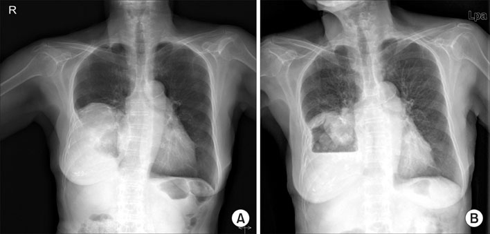
Figure 1 Chest X-ray before 3 months of hospitalization (A) shows a well-defined large mass shadow in the right lower lung field. On admission, chest X-ray taken after massive hemoptysis (B) demonstrates a decreased mass shadow with newly developed air-fluid level which finding is suggestive of intrathoracic bleeding.

Figure 2 Chest CT performed from three months previously (A, B) shows a large ovoid non-enhancing inhomogenous mass with nodular-calcifications in the right posterior and lower pleura, surrounded by egg shell calcification.
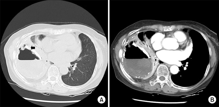
Figure 3 On admission, chest CT (A, B) reveals that there are loss of material and concurrently gain of air-fluid level within the intrathoracic mass-like lesion and bronchopleural fistula (arrow) newly develops in the right lung, which findings verify that some amount of blood in the lesion disappear through the fistulous tract during massive hamoptysis.
Reference
-
1. Reid JD, Kommareddi S, Lankerani M, Park MC. Chronic expanding hematomas. A clinicopathologic entity. JAMA. 1980. 244:2441–2442.2. Kwon YS, Koh WJ, Kim TS, Lee KS, Kim BT, Shim YM. Chronic expanding hematoma of the thorax. Yonsei Med J. 2007. 48:337–340.3. Sato M, Kosaka S, Takahashi T. Life threatening chronic expanding hematoma of the thorax. J Cardiovasc Surg (Torino). 2004. 45:511–514.4. Okubo K, Okamoto T, Isobe J, Ueno Y. Rupture of a chronic expanding hematoma of the thorax into lung parenchyma. J Thorac Cardiovasc Surg. 2004. 127:1838–1840.5. Hanagiri T, Muranaka H, Hashimoto M, Nishio T, Sakai S, Ono M, et al. Chronic expanding hematoma in the chest. Ann Thorac Surg. 1997. 64:559–561.6. Hwang GL, Moffatt SD, Mitchell JD, Leung AN. Chronic expanding hematoma of the thorax. AJR Am J Roentgenol. 2003. 180:1182–1183.7. Athanassiadi K, Reiffen HP, Dickgreber N, Laenger F, Eschenbruch CM, Wilchelmi M, et al. A different surgical approach for an intrathoracic expanding hematoma. J Thorac Cardiovasc Surg. 2007. 133:832–834.8. Labadie EL, Glover D. Physiopathogenesis of subdural hematomas. Part 1: Histological and biochemical comparisons of subcutaneous hematoma in rats with subdural hematoma in man. J Neurosurg. 1976. 45:382–392.
- Full Text Links
-
- Actions
-
Cited
- CITED
-
- Close
- Share
-
- Similar articles
-
- A Case of Spontaneous Chronic Expanding Hematoma in the Thorax
- Use of the Free Flap for Large Defect with Bronchopleural Fistula: Case Report
- Various Bronchial Fistulas: Pitfalls and Usefulness on CT. A Pictorial Review
- Peripheral Bronchopleural Fistula: CT Evaluation in 22 patients
- A Case of Bronchoscopic Treatment of a Bronchopleural Fistula Accompanied by Pneumonia
