Periosteal Fixation in Bilateral Total Third Nerve Palsy
- Affiliations
-
- 1Department of Ophthalmology, Maryknoll Hospital, Pusan, Korea. kris9352@hanmail.net
- KMID: 1476980
- DOI: http://doi.org/10.3341/jkos.2008.49.7.1203
Abstract
-
PURPOSE: We present a new technique of anchoring the eyeball to the nasal periosteum with supramaximal recession of the lateral rectus muscle in one eye for exotropia management in bilateral total third nerve palsy combined with trochlear nerve palsy.
CASE SUMMARY
A 38-year-old man presented with drooping of both upper lids and exodeviation of the left eye with a history of intraventricular hemorrhage 9 months previously. We noted bilateral ptosis, dilated pupils, right fixing eye, left face turn, and left exotropia over 100 prism diopters (PD) in the primary position with an inability to move both eyes together except abduction. He was diagnosed with bilateral total third nerve palsy and trochlear nerve palsy. We fixated the left globe (sclera anterior to the insertion of the medial rectus muscle) to the nasal periosteum including the medial palpebral ligament using a nonabsorbable suture. A large recession of the left lateral rectus muscle (14 mm) was also performed. Ocular alignment in the primary position was exotropia of 25PD and cosmetically satisfactory after 6 months of follow-up.
CONCLUSION
Supramaximal recession of the lateral rectus muscle and periosteal fixation using nonabsorbable suture is an effective technique for the management of exotropia secondary to total third nerve palsy.
Keyword
MeSH Terms
Figure
-

Figure 1. Preoperative photograph in 9 gazes shows bilateral total third nerve palsy and trochlear nerve palsy. The patient had ptosis and mid-dilated fixed pupil in both eyes and left exotropia over 100PD in the primary position. Both eyes had limitation in adduction, elevation, and depression.
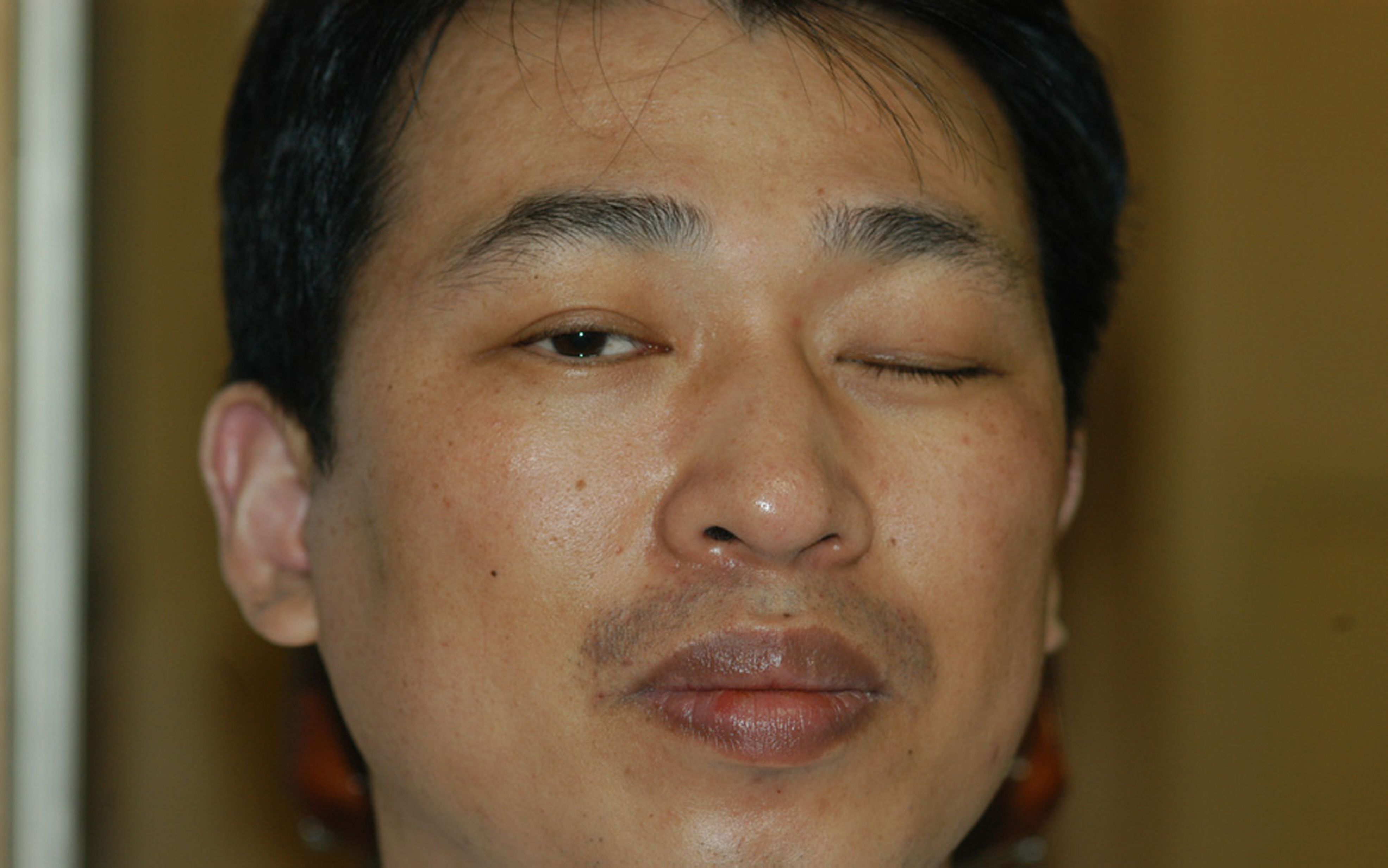
Figure 2. The patient had left face turn and he fixated with the right eye.

Figure 3. Schematic drawings. (A) The anterior lacrimal crest and the insertion of the medial palpebral ligament were exposed. (B) Double armed 5-0 Ethibond suture was passed through the medial palpebral ligament insertion at the anterior lacrimal crest including the periosteum. (Insert) Second bite was taken just lateral to the first. (C) A small buttonhole was created in the medial orbital fascia up to the exposed periosteum by blunt scissors. (D) Ends of the 5-0 Ethibond were brought out into the subtenon space (toward the conjunctival side) using artery clamps. (E) The sutures were secured to the sclera anterior to the medial rectus insertion.

Figure 4. The postoperative 6 months photograph shows left exotropia of 25 PD without hypotropia in the primary position. Mild fullness and congestion over the medial rectus muscle area was observed.
Cited by 2 articles
-
Fixation of the Eyeball to the Periosteum Over the Posterior Lacrimal Crest in Inveterate Exotropia
Bo Ram Seol, Sang In Khwarg, Seong-Joon Kim
J Korean Ophthalmol Soc. 2014;55(3):408-415. doi: 10.3341/jkos.2014.55.3.408.Periosteal Fixation Applied to Patients with Large-angle Paralytic Strabismus
Hee Dong Eom, Young Ki Kwon, Byeong Jae Son, Bo Young Chun
J Korean Ophthalmol Soc. 2018;59(3):268-275. doi: 10.3341/jkos.2018.59.3.268.
Reference
-
References
1. Von Noorden GK. Binocular vision and ocular motility. 3rd ed. St. Louis: CV Mosby;1985. p. 369.2. Rosenbaum AI, Santiago AP. Clinical strabismus management principles and surgical thechnique. Philadelphia: WB Saunders;1999. p. 252.3. Von Noorden GK.Binocular vision and ocular motility. 5th ed. St. Louis: CV Mosby;1996. p. 422.4. Scott AB. Transposition of the superior oblique. Am Orthopt J. 1977; 27:114.
Article5. Saunders RA, Rogers GL. Superior oblique transposition for third nerve palsy. Ophthalmology. 1982; 89:310–6.
Article6. Young TL, Conahan BM, Summers CG, Egbert JE. Anterior transposition of the superior oblique tendon in the treatment of oculomotor nerve palsy and its influence on postoperative hypertropia. J Pediatr Ophthalmol Strabismus. 2000; 37:149–55.
Article7. Helveston EM. Muscle transposition procedures. Surv Ophthalmol. 1972; 16:92–7.8. Helveston EM. Atlas of Strabismus Surgery. 3rd ed. St. Louis: CV Mosby;1985; 254–9.9. Peter LC. The use of the superior oblique as an internal rotator in third nerve paralysis. Trans Am Ophthalmol. 1933; 31:232–7.10. Jackson E. Surgery of the Eye. 3rd ed. Vol. 1. New York: Grune & Stratton;1952; 405.11. Ham DI, Yu YS. Strabismus surgery on congenital oculomotor nerve palsied eye. J Korean Ophthalmol Soc. 1991; 32:262–7.12. Saunders RA, Rogers GL. Superior oblique transposition for third nerve palsy. Ophthalmology. 1982; 89:310–6.
Article13. Salazar-León JA, Ramírez-Ortíz MA, Salas-Vargas M. The surgical correction of paralytic strabismus using fascia lata. J Pediatr Ophthalmol Strabismus. 1998; 35:27–32.
Article14. Srivastava KK, Sundaresh K, Vijayalakshmi P. A new surgical technique for ocular fixation in congenital third nerve palsy. J AAPOS. 2004; 8:371–7.
Article15. Sharma P, Gogoi M, Kedar S, Bhola R. Periosteal fixation in third-nerve palsy. J AAPOS. 2006; 10:324–7.
Article16. Saxena R, Sinha A, Sharma P. . Precaruncular periosteal anchor of medial rectus, a new technique in the management of complete external third nerve palsy. Orbit. 2006; 25:205–8.
Article
- Full Text Links
-
- Actions
-
Cited
- CITED
-
- Close
- Share
-
- Similar articles
-
- A Case of Traumatic Bilateral Abducens Nerve Palsy Associated with Skull Base Fracture
- Periosteal Fixation Applied to Patients with Large-angle Paralytic Strabismus
- Bilateral Traumatic Abducens Nerve Palsy Associated with Hangman's Fracture: Case Report
- Delayed Bilateral Abducens Nerve Palsy after Head Trauma
- Bilateral Simultaneous Bell's Palsy-Two Case Studies
