Cup-to-Disc Ratio, Intraocular Pressure, and Occlusion Site in Branch Retinal Vein Occlusion
- Affiliations
-
- 1Department of Ophthalmology, Chungbuk National University College of Medicine, Cheongju, Korea. simple521@chungbuk.ac.kr
- KMID: 1476963
- DOI: http://doi.org/10.3341/jkos.2007.49.7.1094
Abstract
-
PURPOSE: To investigate correlations among the cup-to-disc ratio (CDR), intraocular pressure (IOP), and the occlusion site in branch retinal vein occlusion (BRVO).
METHODS
This prospective study involved 62 eyes with a diagnosis of BRVO. Fundus photography, fluorescein angiography, Goldmann applanation tonometry, and optical coherence tomography were performed. Correlations among CDR, IOP, and the occlusion site were analyzed.
RESULTS
A negative correlation was found between the occlusion site and IOP (p<0.001, Pearson's correlation analysis) and between the occlusion site and CDR (p<0.001, Pearson's correlation analysis). However, the correlation between IOP and CDR was poor (p=0.092, Pearson's correlation analysis).
CONCLUSIONS
BRVOs with an occlusion site near the optic disc are associated with raised IOP and CDR values. This study suggests that the occurrence of BRVO with an occlusion site near the optic disc indicates that the patient should be evaluated for glaucoma.
Keyword
MeSH Terms
Figure
-
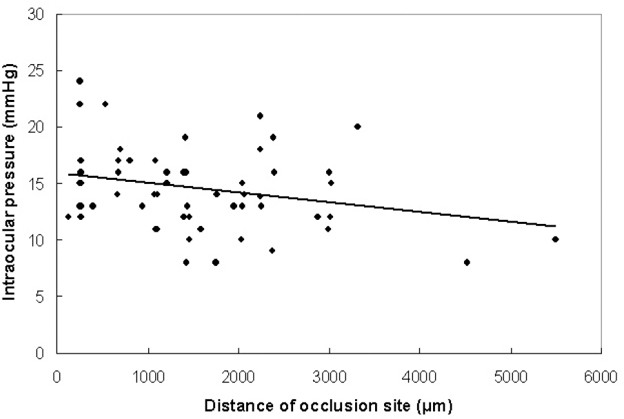
Figure 1. Scatterplot of the correlation between intraocular pressure and occlusion site in eyes with a branch retinal vein occlusion. The correlation was significant (p<0.001).
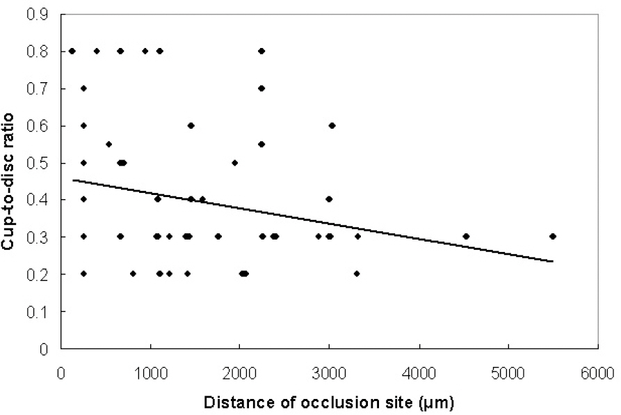
Figure 2. Scatterplot of the correlation between cup-to-disc ratio and occlusion site in eyes with branch retinal vein occlusion. The correlation was significant (p<0.001).

Figure 3. Scatterplot of the correlation between intraocular pressure and cup-to-disc ratio in eyes with branch retinal vein occlusion. No significant correlation was found (p=0.092).
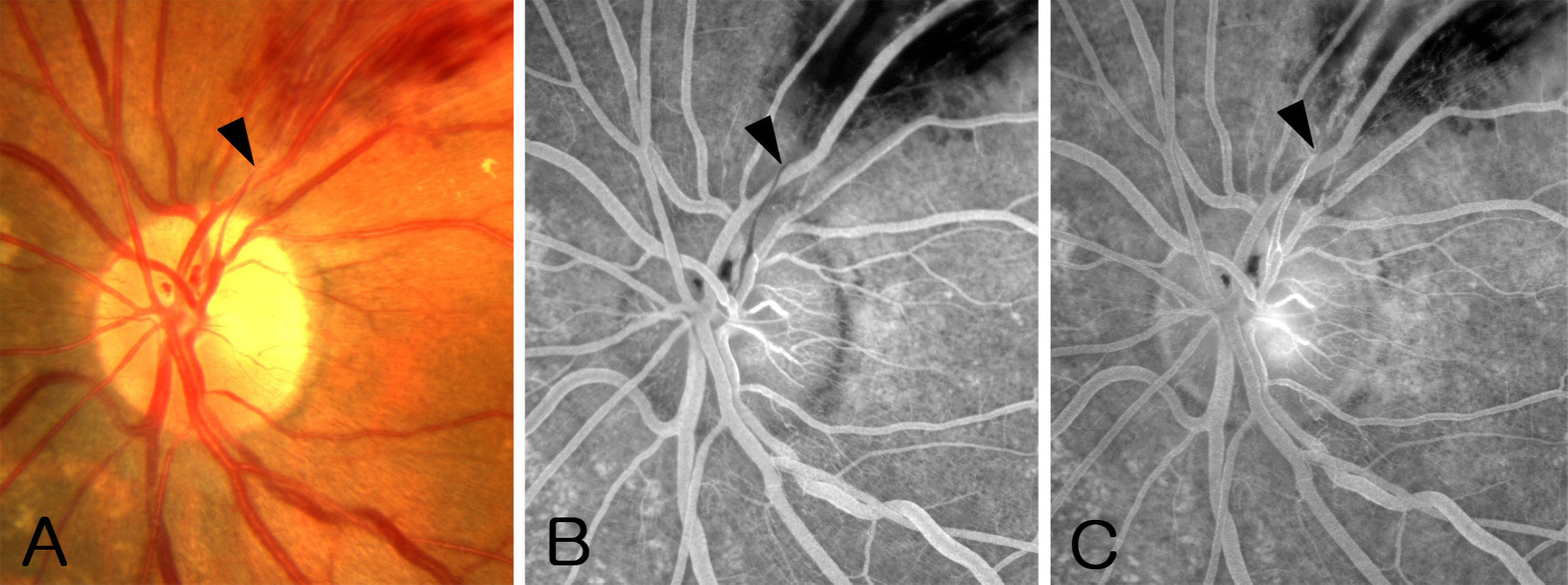
Figure 4. Left eye of a 47-year-old man showing branch retinal vein occlusion. (A) The superior temporal vein was occluded 325 μ m above the disc border (arrowhead), and the proximal portion of the occluded vein was significantly thinner than the distal portion. (A, B) Early and late phase angiogram showing delayed filling and staining of the occluded vein.

Figure 5. Right eye of a 62-year-old man without glaucomatous disc change. (A) The superior temporal branch retinal vein was occluded 3120 µm above the disc border (arrowhead), and the occlusion site was at an artery-vein crossing. (B, C) Early and late phase angiograms showing an abrupt venous caliber change at the occlusion site.

Figure 6. Left eye of a 52-year-old man showing superior temporal branch retinal vein occlusion. The occlusion was occurred on the disc surface (arrowhead), and was more clearly observed in angiograms. The occlusion site was associated with an artery-vein crossing.

Figure 7. Left eye of a 44-year-old woman with glaucomatous disc change and deep excavation. (A) The occlusion occurred in the optic cup (arrowhead). Note the thin collateral vessel along the temporal disc border connecting the superior and inferior retinal vein (arrow). Disc surface hemorrhage was also observed. (B, C) These findings were more clearly observed in the angiograms. The occlusion site was remote from the artery-vein crossing.
Reference
-
References
1. Soni KG, Woodhouse DF. Retinal vascular occlusion as a presenting feature of glaucoma simplex. Br J Ophthalmol. 1971; 55:192–5.
Article2. Frucht J, Shapiro A, Merin S. Intraocular pressure in retinal vein occlusion. Br J Ophthalmol. 1984; 68:26–8.
Article3. Appiah AP, Trempe CL. Risk factors associated with branch vs. central retinal vein occlusion. Ann Ophthalmol. 1989; 21:153–5.4. Risk factors for central retinal vein occlusion The Eye Disease Case-control Study Group. Arch Ophthalmol. 1996; 114:545–54.5. Luntz MH, Schenker HI. Retinal vascular accidents in glaucoma and ocular hypertension. Surv Ophthalmol. 1980; 25:163–7.
Article6. Johnston RL, Brucker AJ, Steinmann W. . Risk factors of branch retinal vein occlusion. Arch Ophthalmol. 1985; 103:1831–2.
Article7. Risk factors for branch retinal vein occlusion. The Eye Disease Case-control Study Group. Am J Ophthalmol. 1993; 116:286–96.8. Lindblom B. Open angle glaucoma and non-central retinal vein occlusion– the chicken or the egg? Acta Ophthalmol Scand. 1998; 76:329–33.9. Beaumont P, Goldberg I, Hollows FC. Optic cup vein occlusion; description of a new entity. Trans Ophthalmol Soc N Z. 1976; 28:115–7.10. Beaumont PE, Kang HK. Cup-to-disc ratio, intraocular pressure, and primary open-angle glaucoma in retinal venous occlusion. Ophthalmology. 2002; 109:282–6.11. Ravalico G, Battaglia Parodi M. Cup/disk ratio in branch retinal vein occlusion. Ophthalmologica. 1991; 203:53–6.12. Hitchings RA, Spaeth GL. Chronic retinal vein occlusion in glaucoma. Br J Ophthalmol. 1976; 60:694–9.
Article13. Klein BE, Magli YL, Richie KA. . Quantitation of optic disc cupping. Ophthalmology. 1985; 92:1654–6.
Article14. Clemett RS. Retinal branch vein occlusion Changes at the site of obstruction. Br J Ophthalmol. 1974; 58:548–54.
Article15. Klein BE, Meuer SM, Knudtson MD, Klein R. The relationship of optic disk cupping to retinal vein occlusion: the Beaver Dam Eye Study. Am J Ophthalmol. 2006; 141:859–62.
Article16. Clemett RS, Kohner EM, Hamilton AM. The visual prognosis in retinal branch vein occlusion. Trans Ophthalmol Soc U K. 1973; 93:523–35.17. Vannas S, Tarkkanen A. Retinal vein occlusion and glaucoma Tonographic study of the incidence of glaucoma and its prognostic significance. Br J Ophthalmol. 1960; 44:583–9.18. Appiah AP, Trempe CL. Differences in contributory factors among hemicentral, central and branch vein occlusions. Ophthalmology. 1989; 96:364–6.19. Bertelsen T. The relationship between thrombosis in the retinal veins and primary glaucoma. Acta Ophthalmol (Copenh). 1961; 39:603–13.
Article20. Clements DB, Elsby JM, Smith WD. Retinal vein occlusion A comparative study of factors affecting the prognosis, including therapeutic trial of Atromid S in this condition. Br J Ophthalmol. 1968; 52:111–6.21. Luntz MH, Schenker HI. Retinal vascular accidents in glaucoma and ocular hypertension. Surv Ophthalmol. 1980; 25:163–7.
Article22. Dobree JH. Venous obstruction and neovascularization at the disc in chronic glaucoma. Trans Opthal Soc U K. 1957; 77:229–37.23. Wise GN, Dollery CT, Henkind P.The retinal circulation. New York: Harper and Row;1971:p. 290–321.24. Sonnsjo B, Krakau CE. Arguments for a vascular glaucoma etiology. Acta Ophthalmol (Copenh). 1993; 71:433–44.
- Full Text Links
-
- Actions
-
Cited
- CITED
-
- Close
- Share
-
- Similar articles
-
- Clinical Features According to the Occlusion Site in Patients with Branch Retinal Vein Occlusion
- Clinical Aspect of Branch Retinal Vein Occlusion
- Retinal Arteriolar Changes in a Patient with Branch Retinal Vein Occlusion
- Clinical observation of the bilateral branch vein occlusion
- Monocytes and High-density Lipoprotein Cholesterol in Branch Retinal Vein Occlusion
