Bowel Entrapment by Fragments of Acetabular Fracture: A Case Report
- Affiliations
-
- 1Department of Orthopaedic Surgery, Asan Medical Center, College of Medicine, University of Ulsan, Seoul, Korea. jjkim2@amc.seoul.kr
- 2Department of Surgery, Asan Medical Center, College of Medicine, University of Ulsan, Seoul, Korea.
- 3Department of Radiology and Research Institute of Radiology, Asan Medical Center, College of Medicine, University of Ulsan, Seoul, Korea.
- 4Department of Orthopaedic Surgery, Haeundae Paik Hospital, College of Medicine, Inje University, Busan, Korea.
- KMID: 1461494
- DOI: http://doi.org/10.12671/jkfs.2010.23.4.373
Abstract
- Abdominal injuries are common in patients with pelvic or acetabular fracture. However intestinal entrapment or perforation caused by fragments of a pelvic or acetabular fracture is rare and to date there has been no report of this occurring in Korea so far. As it is difficult to diagnose intestinal entrapment caused by fragments of pelvic or acetabular fracture, the entrapment therefore results in intestinal perforation, sepsis, and a high mortality rate in the absence of early detection. We present a case of intestinal entrapment and perforation caused by fragments of acetabular fracture as well as a literature review.
Keyword
Figure
-
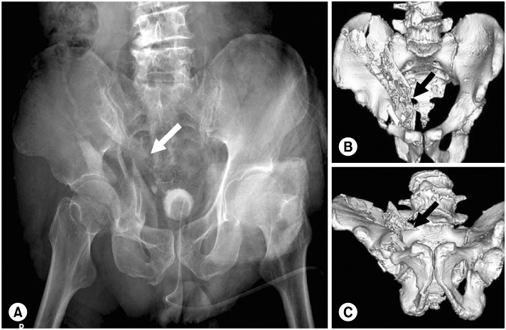
Fig. 1 (A) Initial Pelvis AP view, (B, C) Initial 3D pelvis CT images show both column acetabular fracture with central dislocation. The arrows indicate the fragment protruded medially and superiorly.
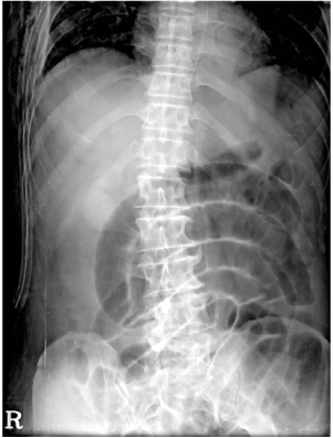
Fig. 2 Supine abdomen x-ray shows intestinal dilatation.
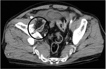
Fig. 3 Initial axial image of pelvis CT shows entrapped bowel between fragments of acetabulum (circle).
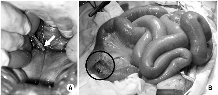
Fig. 4 (A) Acetabular fragment penentrates retroperitoneum and compress small bowel. (B) The distal ileum (circle) is perforated.
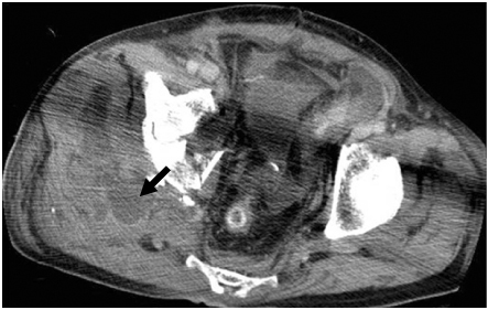
Fig. 5 Pelvis CT image 4 days after reanastomosis operation shows fluid collection (arrow) around gluteus muscle.

Fig. 6 The 6 months follow up Pelvis AP radiograph shows that acetabular fracture doesn't have union and femoral head migrates medially.
Reference
-
1. Arnold GJ. A case of fracture of the pelvis from slight violence, with nipping of small intestine between the fragments causing acute intestinal obstruction and general peritonitis. Lancet. 1909. 27:1157–1158.2. Ashai F, Mam MK, Iqbal S. Ileal entrapment as a complication of fractured pelvis. J Trauma. 1988. 28:551–552.3. Bacarese-Hamilton IA, Bhamra M. Small bowel entrapment following acetabular fracture. Injury. 1991. 22:242–244.
Article4. Buchanan JR. Bowel entrapment by pelvic fracture fragments: a case report and review of the literature. Clin Orthop Relat Res. 1980. 147:164–166.5. Catsikis BD, French WM, Norcus G, Brotman S, Smith JL, Harris RD. CT diagnosis of bowel herniation at pelvic fracture site. J Comput Assist Tomogr. 1989. 13:148–149.
Article6. Charnley GJ, Dorrell JH. Small bowel entrapment in an iliac wing fracture. Injury. 1993. 24:627–628.
Article7. Hurt AV, Ochsner JL, Schiller WR. Prolonged ileus after severe pelvic fracture. Am J Surg. 1983. 146:755–757.
Article8. Lin PS, Cavarocchi NC, Comerota AJ, Resnick EJ. Acute bowel entrapment and perforation following operative reduction of pelvic fracture. J Trauma. 1987. 27:684–686.
Article9. Peltier LF. Complications Associated with Fractures of the Pelvis. J Bone Joint Surg Am. 1965. 47:1060–1069.
Article10. Stubbart JR, Merkley M. Bowel entrapment within pelvic fractures: a case report and review of the literature. J Orthop Trauma. 1999. 13:145–148.
Article
- Full Text Links
-
- Actions
-
Cited
- CITED
-
- Close
- Share
-
- Similar articles
-
- Latent Superior Gluteal Artery Injury by Entrapment between the Fragments in Transverse Acetabular Fracture - A Case Report -
- Vesico-Acetabular Fistula and Urolithiasis in the Hip Joint Cavity due to Persistent Bladder Entrapment after Acetabular Fracture
- Surgical Treatment of Acetabular Posterior Wall Fracture with Hip Arthroscopy: A Case Report
- Combined Femoral and Sciatic Nerve Palsy Associated with Acetabular Fracture and Dislocation: A Case Report
- Sural Nerve Entrapment by Fragments of Calcaneal Fracture (A Case Report)
