Changes in occlusal force and occlusal contact area after orthodontic treatment
- Affiliations
-
- 1Department of Orthodontics, Gangnam Severance Hospital, College of Dentistry, Oral Science Research Institute, The Institute of Cranio-facial Deformity, Yonsei University, Korea. khkim@yuhs.ac
- KMID: 1459571
- DOI: http://doi.org/10.4041/kjod.2010.40.3.176
Abstract
OBJECTIVE
This study was performed to evaluate functional changes of occlusion after orthodontic treatment by measuring the occlusal force (OcFr) and occlusal contact area (OcAr), and to compare OcFr and OcAr change according to premolar extractions.
METHODS
Data were obtained from 74 patients who had finished orthodontic treatment using fixed appliance aged between 18 and 40 years. Subjects were divided into groups who had four premolars extractions or non-extraction (Male extraction-16, Male nonextraction-18, Female extraction-19, Female nonextraction-21). All subjects were asked to bite pressure-sensitive sheets into maximum intercuspation with maximum bite force, and OcFr and OcAr were evaluated by measuring the sheet with a CCD camera. Records were taken right after debonding, 1 week, 1 month, 3 months, 6 months and 1 year after debonding.
RESULTS
OcFr and OcAr increased gradually in all groups during the 1 year retention period (p < 0.05). Male groups showed higher OcFr and OcAr than female groups throughout the retention periods (p < 0.05). There were no statistically significant differences of OcFr and OcAr between extraction and non-extraction groups in both males and females (p > 0.05).
CONCLUSIONS
Occlusion was improved functionally throughout the 1 year retention, and premolar extraction did not induce a decline in the functional aspect of occlusion.
Figure
-
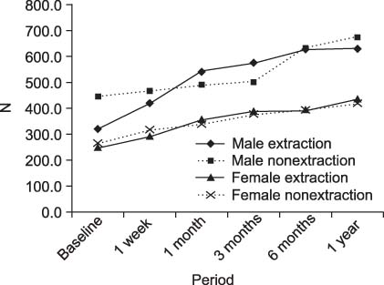
Fig. 1 Occlusal force measured during the 1 year retention period.
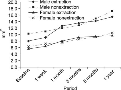
Fig. 2 Occlusal contact area measured during the 1 year retention period.
Cited by 3 articles
-
Differences in molar relationships and occlusal contact areas evaluated from the buccal and lingual aspects using 3-dimensional digital models
Sook-Yoon Jang, Minji Kim, Youn-Sic Chun
Korean J Orthod. 2012;42(4):182-189. doi: 10.4041/kjod.2012.42.4.182.Posterior dental compensation and occlusal function in adults with different sagittal skeletal malocclusions
Soonshin Hwang, Yoon Jeong Choi, Sooin Jung, Sujin Kim, Chooryung J. Chung, Kyung-Ho Kim
Korean J Orthod. 2020;50(2):98-107. doi: 10.4041/kjod.2020.50.2.98.Comparison of the bite force and occlusal contact area of the deviated and non-deviated sides after intraoral vertical ramus osteotomy in skeletal Class III patients with mandibular asymmetry: Two-year follow-up
Hyejin Kwon, Sun-Hyung Park, Hoi-In Jung, Woo-Chan Hwang, Yoon Jeong Choi, Chooryung Chung, Kyung-Ho Kim
Korean J Orthod. 2022;52(3):172-181. doi: 10.4041/kjod21.236..
Reference
-
1. Durbin DS, Sadowsky C. Changes in tooth contacts following orthodontic treatment. Am J Orthod Dentofacial Orthop. 1986. 90:375–382.
Article2. Gazit E, Lieberman MA. Occlusal contacts following orthodontic treatment. Measured by a photocclusion technique. Angle Orthod. 1985. 55:316–320.3. Brudevold F. A basic study of the chewing forces of a denture wearer. J Amer Dental Assoc. 1951. 43:45–51.
Article4. Anderson DJ. Measurement of stress in mastication. II. J Dent Res. 1956. 35:671–673.
Article5. Linderholm H, Wennström A. Isometric bite force and its relation to general muscle forge and body build. Acta Odontol Scand. 1970. 28:679–689.
Article6. Fløystrand F, Kleven E, Øilo G. A novel miniature bite force recorder and its clinical application. Acta Odontol Scand. 1982. 40:209–214.
Article7. Gibbs CH, Mahan PE, Lundeen HC, Brehnan K, Walsh EK, Holbrook WB. Occlusal forces during chewing and swallowing as measured by sound transmission. J Prosthet Dent. 1981. 46:443–449.
Article8. Hidaka O, Iwasaki M, Saito M, Morimoto T. Influence of clenching intensity on bite force balance, occlusal contact area, and average bite pressure. J Dent Res. 1999. 78:1336–1344.
Article9. Ando K, Kurosawa M, Fuwa Y, Kondo T, Goto S. A study on measuring occlusal contact area using silicone impression materials: an application of this method to the bite force measurement system using the pressure-sensitive sheet. Dent Mater J. 2007. 26:898–905.
Article10. Kwon HK, Yoo JH, Kwon YS, Kim BI. Comparison of bite force with dental prescale and unilateral bite force recorder in healthy subjects. J Korean Acad Prosthodont. 2006. 44:103–111.11. Hassan SG, Yamada K, Rakiba S, Morita S, Hanada K. Relationship between craniofacial morphology and occlusal force in adults with normal occlusion. J Jpn Orthod Soc. 1997. 56:348–361.12. Hattori Y, Satoh C, Watanabe M. Bite force distribution on dental arch during clenching. J Jpn Soc Stomatognathic Func. 1996. 2:111–117.
Article13. Sultana MH, Yamada K, Hanada K. Changes in occlusal force and occlusal contact area after active orthodontic treatment: a pilot study using pressure-sensitive sheets. J Oral Rehabil. 2002. 29:484–491.
Article14. Noguchi T, Fukuda M, Tauchi S, Kinoshita S. Wrapping of occlusal prescale for occlusal examination. Nippon Shishubyo Gakkai Kaishi. 1983. 25:575–581.15. Suzuki T, Watanabe T, Yoshitomi N, Ishinaba S, Kumagai H, Uchida T, et al. Evaluation of a new measuring system for occlusal force with pressure sensitive sheet. J Jpn Prosthodont Soc. 1994. 38:966–973.
Article16. Matsui Y, Ohno K, Michi K, Suzuki Y, Yamagata K. A computerized method for evaluating balance of occlusal load. J Oral Rehabil. 1996. 23:530–535.
Article17. Bachus KN, DeMarco AL, Judd KT, Horwitz DS, Brodke DS. Measuring contact area, force, and pressure for bioengineering applications: using Fuji Film and TekScan systems. Med Eng Phys. 2006. 28:483–488.
Article18. Braun S, Bantleon HP, Hnat WP, Freudenthaler JW, Marcotte MR, Johnson BE. A study of bite force, part 1: Relationship to various physical characteristics. Angle Orthod. 1995. 65:367–372.19. Ingervall B, Minder C. Correlation between maximum bite force and facial morphology in children. Angle Orthod. 1997. 67:415–424.20. Helkimo E, Carlsson GE, Helkimo M. Bite force and state of dentition. Acta Odontol Scand. 1977. 35:297–303.
Article21. Yoon HR. The comparison of occlusal force according to occlusion, skeletal pattern, age and gender in Koreans. 2010. Yonsei University;Master. thesis.22. Cha BK, Kim CH, Baek SH. Skeletal sagittal and vertical facial types and electromyographic activity of the masticatory muscle. Angle Orthod. 2007. 77:463–470.
Article23. Miralles R, Hevia R, Contreras L, Carvajal R, Bull R, Manns A. Patterns of electromyographic activity in subjects with different skeletal facial types. Angle Orthod. 1991. 61:277–284.24. Fogle LL, Glaros AG. Contributions of facial morphology, age, and gender to EMG activity under biting and resting conditions: a canonical correlation analysis. J Dent Res. 1995. 74:1496–1500.
Article25. Ringqvist M. Isometric bite force and its relation to dimensions of the facial skeleton. Acta Odontol Scand. 1973. 31:35–42.
Article26. Proffit WR, Fields HW, Nixon WL. Occlusal forces in normal- and long-face adults. J Dent Res. 1983. 62:566–570.
Article27. Nakajima A, Watanabe K, Sugi E, Yasuda K, Maruyama J, Ono S, et al. Changes in occlusal force and occlusal contact area during retention - consideration form three cases. Nihon Univ Dent J. 1994. 68:457.28. Razdolsky Y, Sadowsky C, BeGole EA. Occlusal contacts following orthodontic treatment: a follow-up study. Angle Orthod. 1989. 59:181–185.29. Bakke M. Mandibular elevator muscles: physiology, action, and effect of dental occlusion. Scand J Dent Res. 1993. 101:314–331.
Article
- Full Text Links
-
- Actions
-
Cited
- CITED
-
- Close
- Share
-
- Similar articles
-
- Comparisons of occlusal force according to occlusal relationship, skeletal pattern, age and gender in Koreans
- Considerations in the reliability of occlusal indicators and occlusal contact marks
- Occlusal force evaluation and its clinical implication in dental clinics
- Effectiveness of clinical remounting improving balanced occlusion of complete dentures
- Changes of bite force and dynamic functional occlusion analysis after occlusal stabilization splint therapy in sleep bruxism patients: a pilot study
