Assessment of antero-posterior skeletal relationships in adult Korean patients in the natural head position and centric relation
- Affiliations
-
- 1Associate Professor, Department of Orthodontics, Kangnam Sacred Heart Hospital, Hallym University, Korea.
- 2Assistant Professor, Department of Preventive Dentistry, School of Dentistry, Seoul National University, Korea.
- 3Professor, Department of Oral and Maxillofacial Surgery, Kangnam Sacred Heart Hospital, Hallym University, Korea.
- 4Chairman, Department of Orthodontics, Chong-A Dental Hospital, Korea.
- 5Assistant Professor, Department of Oral and Maxillofacial Surgery, Kangnam Sacred Heart Hospital, Hallym University, Korea.
- 6Assistant Professor, Department of Orthodontics, Hangang Sacred Heart Hospital, Hallym University, Korea. aurora0@hanmail.net.
- KMID: 1459544
- DOI: http://doi.org/10.4041/kjod.2010.40.6.421
Abstract
OBJECTIVE
This study aimed to verify the intra-individual reproducibility of the natural head position (NHP) in adult Korean patients in the centric relation (CR) position and to prove the inter-individual variability of the Frankfurt horizontal (FH) plane and sella-nasion (SN) line compared to the true horizontal line (THL). In addition, the study aimed to investigate the correlations between linear measurements from A-point and B-point to the nasion true vertical line (NTVL) and angular measurements from A-point and B-point to the SN line.
METHODS
Two lateral cephalograms were taken of 116 subjects (23 males, 93 females) with CR wax bites in a NHP at a one-week interval.
RESULTS
Method errors of three variables and intraclass correlation coefficients of six parameters proved the intra-individual reproducibility of NHP (p < 0.001). The angle of the FH to the THL was not significantly different from 0degrees (p > 0.05), but it was clinically variable (SD 3.89degrees) on the inter-individual level. Conversely, the angle of the SN line to the THL was significantly different from 7degrees (p < 0.05). Very low correlation was found between the linear measurements and angular measurements of A-point and B-point (p < 0.01).
CONCLUSIONS
The NTVL could be a useful reference line for assessing the antero-posterior position of the maxilla and mandibleof Korean adult patients in NHP and CR.
Keyword
Figure
-

Fig 1. Registration of the centric relation wax bite. A, Guiding the mandible using the tripoding method; B, centric relation wax bite (anterior and posterior portion).

Fig 2. Registration in the natural head position and lateral cephalogram. A, Subjects looked into their eyes in the mirror (orthoposition); B, standing position with arms and chin relaxed.
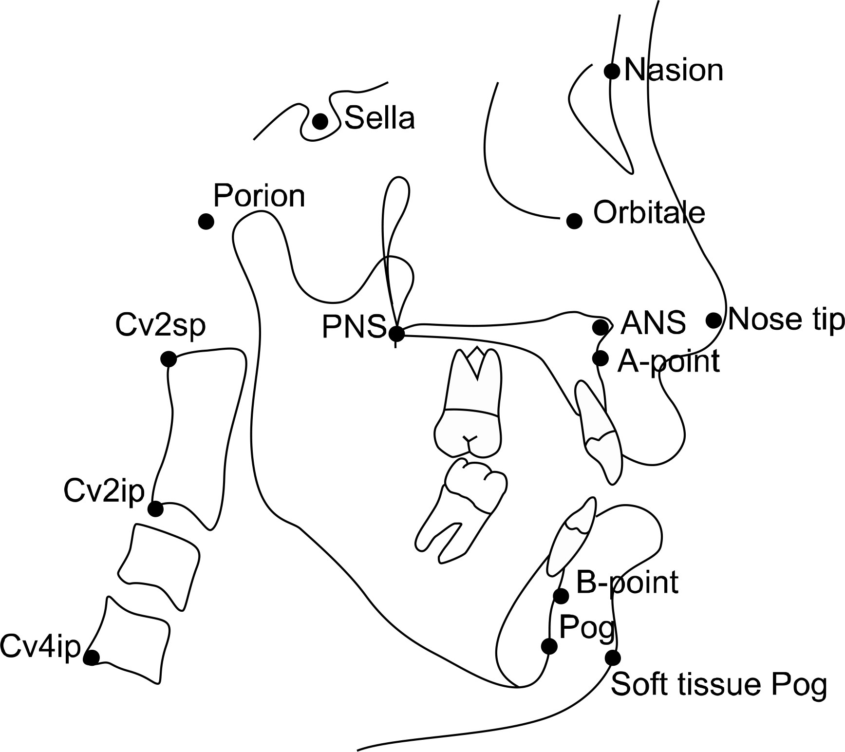
Fig 3. Reference points used in this study.

Fig 4. Reference planes used in this study. 1, Nasion true vertical line (NTVL) passing through the nasion point perpendicular to the floor; 2, E-line, tip of nose-soft tissue pogonion; 3, cervical vertebrae tangent-plane the posterior tangent to the odontoid process through cv4ip; 4, odontoid process tangent plane, the posterior tangent to the odontoid process through cv2ip; 5, palatal plane, ANS-PNS; 6, true horizontal line passing through nasion point perpendicular to the NTVL; 7, sella-nasion plane; 8, Frankfurt horizontal plane, porion-orbitale.

Fig 5. Angular measurements. A, 1, Nasion true vertical line (NTVL) to palatal plane; 2, NTVL to E-Line; 3, NTVL to cervical vertebrae tangent; 4, NTVL to sella-nasion (SN); 5, true horizontal line (THL) to odontoid process tangent (OPT); 6, SN to OPT. B, 1, THL to Frankfurt horizontal plane; 2, THL to SN.

Fig 6. Linear measurements. A, 1, Nasion true vertical line (NTVL to A-point); 2, NTVL to B-point. B, 1, Sella nasion A-point (SNA) angle; 2, sella nasion B-point (SNB) angle.
Reference
-
1.Broadbent BH. A new X-ray technique and its application to orthodontia. Angle Orthod. 1931. 1:45–66.2.Hofrath H. Die Bedeutung der Röntgenfern –und Abstand-saufnahme für die Diagnostik der kieferanomalien. Fortschritte der kieferorthopädie. 1931. 1:232–58.
Article3.Downs WB. Variations in facial relationships; their significance in treatment and prognosis. Am J Orthod. 1948. 34:812–40.
Article4.Steiner CC. Cephalometrics for you and me. Am J Orthod. 1953. 39:729–55.
Article5.Riedel RA. Relation of maxillary structures to cranium in malocclusion and in normal occlusion. Angle Orthod. 1952. 22:142–45.6.Tweed CH. The Frankfort-mandibular plane angle in orthodontic diagnosis classification, treatment planning, and prognosis. Am J Orthod Oral Surg. 1946. 32:175–230.
Article7.Tweed CH. The Frankfort-mandibular incisor angle (FMIA) in orthodontic diagnosis, treatment planning and prognosis. Angle Orthod. 1954. 24:121–69.8.Ricketts RM. Cephalometric synthesis: An exercise in stating objectives and planning treatment with tracings of the head roentgenogram. Am J Orthod. 1960. 46:647–73.9.Jenkins DH. Analysis of orthodontic deformity employing lateral cephalostatic radiography. Am J Orthod. 1955. 41:442–52.
Article10.Jacobson A. The “Wits” appraisal of jaw disharmony. Am J Orthod. 1975. 67:125–38.
Article11.McNamara JA Jr. A method of cephalometric evaluation. Am J Orthod. 1984. 86:449–69.
Article12.Taylor CM. Changes in the relationship of nasion, point A, and point B and the effect upon ANB. Am J Orthod. 1969. 56:143–63.13.Beatty EJ. A modified technique for evaluating apical base relationships. Am J Orthod. 1975. 68:303–15.
Article14.Broca M. Sur les projections de la tète, et sur un nouveau pro-cède de cephalometrié. Bull de la SociétéD'Anthropologie de Paris. 1862. 3:514–44.15.Moorrees CFA., Kean MR. Natural head position: a basic consideration in the interpretation of cephalometric radiographs. Am J Phys Anthrop. 1958. 16:213–34.
Article16.Leitão P., Nanda RS. Relationship of natural head position to craniofacial morphology. Am J Orthod Dentofacial Orthop. 2000. 117:406–17.17.Peng L., Cooke MS. Fifteen-year reproducibility of natural head posture: A longitudinal study. Am J Orthod Dentofacial Orthop. 1999. 116:82–5.
Article18.Choi BT. Articulator mounting. In: Choi BT editor. Steps of preparation for orthognathic surgery. Seoul: Jee sung;2004. p. 113–32.19.Solow B., Tallgren A. Natural head position in standing subjects. Acta Odontol Scand. 1971. 29:591–607.
Article20.Dahlberg G. Statistical methods for medical and biological students. New York: Interscience Publications;1940.21.Park EJ., Suhr CH. Study of craniocervical posture and craniofacial morphology in Korean young adults. Korean J Orthod. 1995. 25:129–42.22.Lee CM., Cha KS. A study on the adaptation of the craniofacial structure to the variations of head postures. Korean J Orthod. 1992. 22:169–77.23.Kim HR., Lee DY., Kim KW., Yoon YJ. A study on the reproducibility of the natural head position according to the skeletal malocclusion types and sex. Korean J Orthod. 2000. 30:307–15.24.Hanau RL. Occlusal changes in centric relation. J Am Dent Assoc. 1929. 16:1903–15.25.The glossary of prosthodontic terms. The Academy of Prosthodontics. J Prosthet Dent. 1994. 71:41–112.26.Lundeen HC., Shryock EF., Gibbs CH. An evaluation of mandibular border movements: their character and significance. J Prosthet Dent. 1978. 40:442–52.
Article27.Lucia VO. A technique for recording centric relation. J Prosthet Dent. 1964. 14:492–505.
Article28.Dawson PE. Centric relation. In: Dawson PE editor. Evaluation, diagnosis, and treatment of occlusal problem. St Loius: CV Mosby;1974. p. 48–70.29.Strohaver RA. A comparison of articulator mountings made with centric relation and myocentric position records. J Prosthet Dent. 1972. 28:379–90.
Article30.Long JH. Locating centric relation with a leaf gauge. J Prosthet Dent. 1973. 29:608–10.
Article31.Dawson PE. Optimum TMJ condyle position in clinical practice. Int J Periodontics Restorative Dent. 1985. 5:10–31.32.Houston WJ. The analysis of errors in orthodontic measurements. Am J Orthod. 1983. 83:382–90.
Article33.Huggare JA. A natural head position technique for radiographic cephalometry. Dentomaxillofac Radiol. 1993. 22:74–6.
Article34.Cooke MS., Wei SH. The reproducibility of natural head posture: a methodological study. Am J Orthod Dentofacial Orthop. 1988. 93:280–8.
Article35.Showfety KJ., Vig PS., Matteson S. A single method for taking natural head position cephalograms. Am J Orthod. 1983. 83:495–500.36.Bjerin R. A comparison between the Frankfort Horizontal and the sella turcica-nasion line as reference planes in cephalometric analysis. Acta Odontologica Scandinavica. 1957. 15:1–12.37.Ferrario VF., Sforza C., Germanò D., Dalloca LL., Miani A Jr. Head posture and cephalometric analyses: an integrated photo-graphic/radiographic technique. Am J Orthod Dentofacial Orthop. 1994. 106:257–64.
Article38.Raju NS., Prasad KG., Jayade VP. A modified approach for obtaining cephalograms in the natural head position. J Orthod. 2001. 28:25–8.
Article39.Uşümez S., Orhan M. Inclinometer method for recording and transferring natural head position in cephalometrics. Am J Orthod Dentofacial Orthop. 2001. 120:664–70.40.Bister D., Edler RJ., Tom BD., Prevost AT. Natural head posture-considerations of reproducibility. Eur J Orthod. 2002. 24:457–70.
Article41.Madsen DP., Sampson WJ., Townsend GC. Craniofacial reference plane variation and natural head position. Eur J Orthod. 2008. 30:532–40.
Article42.Marcotte MR. Head posture and dentofacial proportions. Angle Orthod. 1981. 51:208–13.43.Lundström F., Lundström A. Natural head position as a basis for cephalometric analysis. Am J Orthod Dentofacial Orthop. 1992. 101:244–7.
Article44.Lundström A., Lundström F. The Frankfort horizontal as a basis for cephalometric analysis. Am J Orthod Dentofacial Orthop. 1995. 107:537–40.
Article45.Cooke MS., Wei SH. A summary five-factor cephalometric analysis based on natural head posture and the true horizontal. Am J Orthod Dentofacial Orthop. 1988. 93:213–23.46.Viazis AD. A cephalometric analysis based on natural head position. J Clin Orthod. 1991. 25:172–81.47.Kang SG., Lee YJ., Park YG. A comparative study of soft tissue profile between Korean and Caucasian young adults under NHP. Korean J Orthod. 2003. 33:323–37.48.Michiels LY., Tourne LP. Nasion true vertical: a proposed method for testing the clinical validity of cephalometric measurements applied to a new cephalometric reference line. Int J Adult Orthodon Orthognath Surg. 1990. 5:43–52.
- Full Text Links
-
- Actions
-
Cited
- CITED
-
- Close
- Share
-
- Similar articles
-
- Relative position of mandibular condyle and comparison of reproducibility utilizing different centric relation record taking methods
- Conserving the centric relation position of condylar head with mini-Hoffmann sets in vertical ramus osteotomy
- A study on the cephalometric changes by the displacement of the mandibular condyles
- A study on the antero-posterior morphology of the upper face in angle's Class III malocclusion patients
- A study on the reproducibility of the natural head position according to the skeletal malocclusion type and sex
