J Cardiovasc Ultrasound.
2010 Mar;18(1):1-5. 10.4250/jcu.2010.18.1.1.
Usefulness of Mitral Annular Systolic Velocity in the Detection of Left Ventricular Systolic Dysfunction: Comparison with Three Dimensional Echocardiographic Data
- Affiliations
-
- 1Division of Cardiology, Department of Internal Medicine, School of Medicine, Chungnam National University, Chungnam National University Hospital, Daejeon, Korea. jaehpark@cnuh.co.kr
- KMID: 1457197
- DOI: http://doi.org/10.4250/jcu.2010.18.1.1
Abstract
- BACKGROUND
Although the modified Simpson's method is widely used for the assessment of left ventricular ejection fraction (LVEF), it has limitations including relatively high inter- and intra-observer variability and time consuming nature. We want to evaluate whether assessing mitral annular systolic velocity (S' velocity) by tissue Doppler imaging (TDI) can be used to evaluate LV systolic function with comparing LVEF by three dimensional echocardiography (3DE). METHODS: We examined 3DE and TDI studies of patients between January and August 2008. 3DE LVEF was measured by offline commercial computer software EchoPac PC(R) (GE, Andover, MA, USA). S' velocity was obtained from the medial side with apical four chamber view by pulsed-wave Doppler with TDI. RESULTS: We included 125 patients (78 males (62.4%), mean age: 57.5+/-13.0 years). The mean S' velocity was 7.7+/-1.9 cm/s and the mean LVEF was 57.2+/-10.4%. The S' velocity measured by TDI showed a linear correlation with LVEF measured by 3DE (r=0.688, p<0.001). Study patients were divided into two groups according to the presence of LV systolic dysfunction: Group I (normal LVEF), n=102 and Group II (LVEF <50%), n=23. For prediction of significant LV systolic dysfunction by the receiver operating characteristic curve according to S' velocity, the optimal cutoff value was 6.8 cm/s. At this cutoff value, the sensitivity and specificity were 94.1% and 87%, respectively. CONCLUSION: In this study, S' velocity measured by TDI showed a significant correlation with three dimensional LVEF and can be used to detect patients with LV systolic dysfunction.
Keyword
MeSH Terms
Figure
-
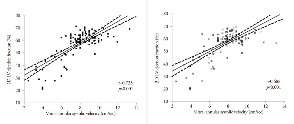
Fig. 1 Correlations between mitral systolic velocity and 2- and 3-dimensional left ventricular ejection fraction.
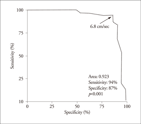
Fig. 2 Receiver operating characteristic (ROC) curve analysis in detecting left ventricular systolic dysfunction using S' velocity. S' velocity lower than 6.8 cm/sec has the best sensitivity and specificity in detection of left ventricular systolic dysfunction.
Reference
-
1. Becker LC, Silverman KJ, Bulkley BH, Kallman CH, Mellits ED, Weisfeldt M. Comparison of early thallium-201 scintigraphy and gated blood pool imaging for predicting mortality in patients with acute myocardial infarction. Circulation. 1983. 67:1272–1282.
Article2. Pilote L, Silberberg J, Lisbona R, Sniderman A. Prognosis in patients with low left ventricular ejection fraction after myocardial infarction. Importance of exercise capacity. Circulation. 1989. 80:1636–1641.
Article3. Pfeffer MA, Braunwald E, Moyé LA, Basta L, Brown EJ Jr, Cuddy TE, Davis BR, Geltman EM, Goldman S, Flaker GC. The SAVE Investigators. Effect of captopril on mortality and morbidity in patients with left ventricular dysfunction after myocardial infarction. Results of the survival and ventricular enlargement trial. N Engl J Med. 1992. 327:669–677.
Article4. Isaaz K, Thompson A, Ethevenot G, Cloez JL, Brembilla B, Pernot C. Doppler echocardiographic measurement of low velocity motion of the left ventricular posterior wall. Am J Cardiol. 1989. 64:66–75.
Article5. Sohn DW, Chai IH, Lee DJ, Kim HC, Kim HS, Oh BH, Lee MM, Park YB, Choi YS, Seo JD, Lee YW. Assessment of mitral annulus velocity by Doppler tissue imaging in the evaluation of left ventricular diastolic function. J Am Coll Cardiol. 1997. 30:474–480.
Article6. Nah DY, Park JH. Mitral annulus velocity measured by pulsed wave Doppler tissue imaging in healthy Korean people. J Korean Soc Echocardiogr. 1999. 7:169–174.
Article7. Bland JM, Altman DG. Statistical methods for assessing agreement between two methods of clinical measurement. Lancet. 1986. 1:307–310.
Article8. Carr KW, Engler RL, Forsythe JR, Johnson AD, Gosink B. Measurement of left ventricular ejection fraction by mechanical cross-sectional echocardiography. Circulation. 1979. 59:1196–1206.
Article9. Schiller NB, Acquatella H, Ports TA, Drew D, Goerke J, Ringertz H, Silverman NH, Brundage B, Botvinick EH, Boswell R, Carlsson E, Parmley WW. Left ventricular volume from paired biplane two-dimensional echocardiography. Circulation. 1979. 60:547–555.
Article10. Doughty RN, Wright S, Whalley GA. Echocardiography or radionuclide methods for assessment of left ventricular function in acute myocardial infarction. Am J Cardiol. 1998. 82:704.11. Nagueh SF. Tissue Doppler imaging for the assessment of left ventricular diastolic function. J Cardiovasc Ultrasound. 2008. 16:76–79.
Article12. Gulati VK, Katz WE, Follansbee WP, Gorcsan J 3rd. Mitral annular descent velocity by tissue Doppler echocardiography as an index of global left ventricular function. Am J Cardiol. 1996. 77:979–984.
Article13. Simonson JS, Schiller NB. Descent of the base of the left ventricle: an echocardiographic index of left ventricular function. J Am Soc Echocardiogr. 1989. 2:25–35.
Article14. Vinereanu D, Nicolaides E, Tweddel AC, Fraser AG. "Pure" diastolic dysfunction is associated with long-axis systolic dysfunction. Implications for the diagnosis and classification of heart failure. Eur J Heart Fail. 2005. 7:820–828.
Article15. Vinereanu D, Khokhar A, Tweddel AC, Cinteza M, Fraser AG. Estimation of global left ventricular function from the velocity of longitudinal shortening. Echocardiography. 2002. 19:177–185.
Article16. Alam M, Wardell J, Andersson E, Nordlander R, Samad B. Assessment of left ventricular function using mitral annular velocities in patients with congestive heart failure with or without the presence of significant mitral regurgitation. J Am Soc Echocardiogr. 2003. 16:240–245.
Article17. Fukuda K, Oki T, Tabata T, Iuchi A, Ito S. Regional left ventricular wall motion abnormalities in myocardial infarction and mitral annular descent velocities studied with pulsed tissue Doppler imaging. J Am Soc Echocardiogr. 1998. 11:841–848.
Article18. Yuda S, Inaba Y, Fujii S, Kokubu N, Yoshioka T, Sakurai S, Nishizato K, Fujii N, Hashimoto A, Uno K, Nakata T, Tsuchihashi K, Miura T, Ura N, Natori H, Shimamoto K. Assessment of left ventricular ejection fraction using long-axis systolic function is independent of image quality: a study of tissue Doppler imaging and m-mode echocardiography. Echocardiography. 2006. 23:846–852.
Article19. Dagianti A, Vitarelli A, Conde Y, Penco M, Fedele F, Dagianti A. Assessement of regional left ventricular function during exercise test with pulsed tissue Doppler imaging. Am J Cardiol. 2000. 86:30G–32G.
Article20. Alam M, Wardell J, Andersson E, Samad BA, Nordlander R. Effects of first myocardial infarction on left ventricular systolic and diastolic function with the use of mitral annular velocity determined by pulsed wave Doppler tissue imaging. J Am Soc Echocardiogr. 2000. 13:343–352.
Article21. Monaghan MJ. Role of real time 3D echocardiography in evaluating the left ventricle. Heart. 2006. 92:131–136.
Article
- Full Text Links
-
- Actions
-
Cited
- CITED
-
- Close
- Share
-
- Similar articles
-
- Comparison of Postoperative LV Function after Mitral Valve Replacement and Predictor of Postoperative LV Function in Chronic Mitral Regurgitation
- Clinical Significance of Atrioventricular Plane Displacement for Evaluating Left Ventricular Diastolic Dysfunction
- Echocardiographic Doppler Mitral Valve Flow Velocity In Hypertension
- Evaluation of Left Ventricular Systolic Function by Tissue Doppler Imaging
- Assessment of Cardiac Function in Neonates by Using Tissue Doppler Imaging
