A modified device for intraoral radiography to assess the distal osseous defects of mandibular second molar after impacted third molar surgery
- Affiliations
-
- 1Stomatology Department, Faculty of Medicine and Dentistry, Santiago de Compostela University, Santiago de Compostela, Spain.
- 2Comprehensive Adult Dental Care, Stomatology Department, Faculty of Medicine and Dentistry, Santiago de Compostela University, Santiago de Compostela, Spain. mercedes.gallas.torreira@usc.es
- KMID: 1449956
- DOI: http://doi.org/10.5624/isd.2011.41.3.115
Abstract
- PURPOSE
This article is to describe a modified device for intraoral radiography which was developed to obtain reproducible radiographic images for assessment of distal osseous defects of the mandibular second molar (2 Mm) after impacted third molar (3 Mm) surgery.
MATERIALS AND METHODS
A commercial available alignment system for posterior region was modified by adding a reference gauge pin (millimetric) and threading a hollow acrylic cylinder at the ring of the radiographic positioner to attach the X-ray collimator. The design included customized resin acrylic stent for the occlusal surface of the 2Mm in maximum intercuspal position, individualizing the biteblock positioner. Periapical radiographs were taken before and after surgical extraction of 3 Mm, employing the radiographic technique of parallelism described by Kugelberg (1986) with this modified film holder and inserting the gauge pin on the deepest bone probing depth point.
RESULTS
This technique permitted to obtain standardized periapical radiographs with a moderate to high resolution, repeatability, and accuracy. There was no difference between the measurements on the pre- and post-operative radiographs. This technique allowed better maintenance of the same geometric position compared with conventional one. The insertion of the gauge pin provided the same reference point and localized the deepest osseous defect on the two-dimensional radiographs.
CONCLUSION
This technique allowed better reproducibility in posterior radiographic records (distal surface of 2 Mm) and more accurate measurements of radiographic bone level by the use of a millimetric pin.
Figure
-
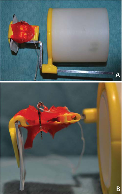
Fig. 1 A. Hollow cylinder adapted to the X-ray film positioner (posterior system Rinn® XCP Instrument Kit, Dentsply, Elgin, IL, USA) with a bite-block customized with acrylic resin (Pattern Resin®, GC America Inc., Alsip, IL, USA) for each patient. B. Detail of the bite-block customized.
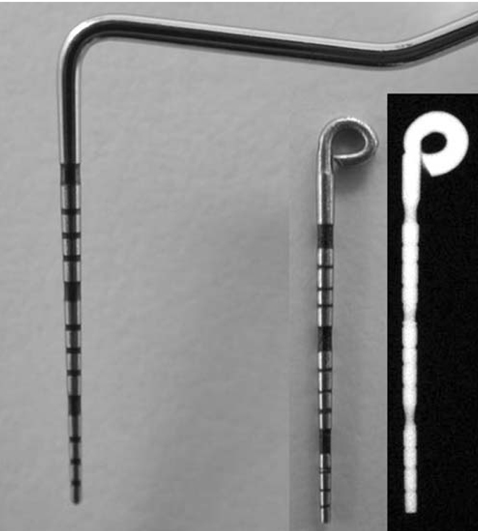
Fig. 2 Gauge pin calibrated-tip of periodontal standardized probe (PCP-UNC15®, Hu-Friedy, Chicago, IL, USA) and radiographic image.
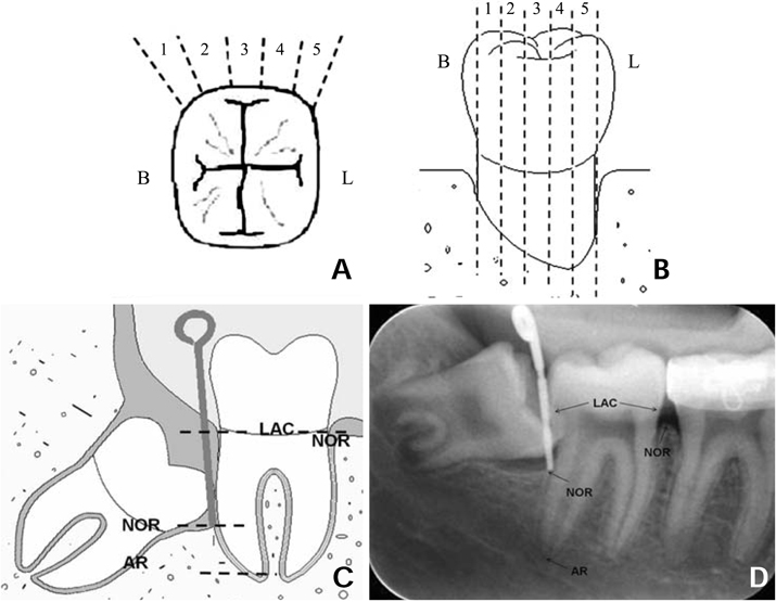
Fig. 3 Division of distal aspect of mandibular second molar. A. Occlusal view. B. Distal view. C. Gauge pin inserted in the deepest probing depth. D. Radiographic image obtained with the anatomic reference points. B; bucal aspect of mandibular second molar, L; lingual aspect of mandibular second molar, AR; radiographic apex of distal root of mandibular second molar, NOR; radiographic bone level, LAC; cementoenamel junction.
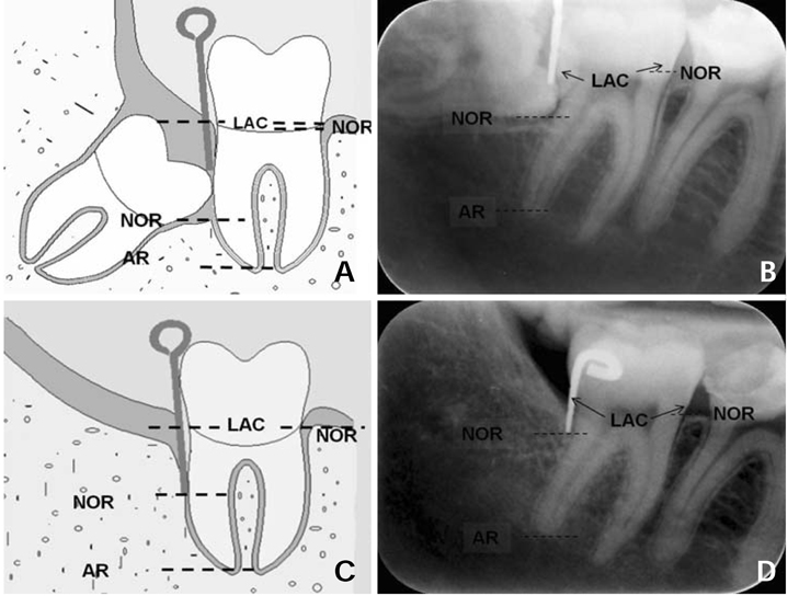
Fig. 4 Drawing (A) and radiographic image (B) prior extraction of the mandibular third molar. Drawing (C) and radiographic image (D) after extraction of the mandibular third molar. AR; radiographic apex of distal root of mandibular second molar, NOR; radiographic bone level, LAC; cementoenamel junction.

Fig. 5 A. Radiograph made only with posterior X-ray film positioner. B. Radiograph made with posterior X-ray film positioner adapted to the hollow cylinder. C. Radiograph made with the hollow cylinder adapted to the posterior X-ray film positioner, with a bite-block customized with acrylic resin. D. Radiograph made with the hollow cylinder adapted to the posterior X-ray film positioner, with a bite-block customized and the gauge pin calibrated inserted in the deepest probing deep.
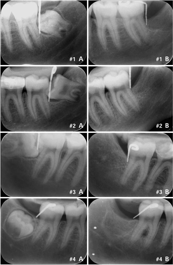
Fig. 6 Four examples of periapical radiographic images. A. Initial radiographic image before extraction of the mandibular third molar. B. Final radiographic image after extraction of the mandibular third molar.
Reference
-
1. White SC, Pharoah MJ. White SC, Pharoah MJ, editors. Intraoral radiographic examinations. Oral radiology; principles and interpretation. 2004. 5th ed. St. Louis: Mosby;121–125.2. Benn DK. A review of the reliability of radiographic measurements in estimating alveolar bone changes. J Clin Periodontol. 1990. 17:14–21.
Article3. Hausmann E. Radiographic and digital imaging in periodontal practice. J Periodontol. 2000. 71:497–503.
Article4. Hausmann E, Allen K, Christersson L, Genco RJ. Effect of x-ray beam vertical angulation on radiographic alveolar crest level measurement. J Periodontal Res. 1989. 24:8–19.
Article5. Jenkins SM, Dummer PM, Newcombe RG. Radiographic amelocemental junction and alveolar crest: effect of X-ray beam angulation. J Oral Rehabil. 1995. 22:679–684.
Article6. Sewerin I, Andersen V, Stoltze K. Influence of projection angles upon position of cementoenamel junction on radiographs. Scand J Dent Res. 1987. 95:74–81.
Article7. Hausmann E, Allen K. Reproducibility of bone height measurements made on serial radiographs. J Periodontol. 1997. 68:839–841.
Article8. Carpio LC, Hausmann E, Dunford RG, Allen KM, Christersson LA. Evaluation of a simple modified radiographic alignment system for routine use. J Periodontol. 1994. 65:62–67.
Article9. Kugelberg CF, Ahlstrom U, Ericson S, Hugoson A. Periodontal healing after impacted lower third molar surgery. Precision and accuracy of radiographic assessment of intrabony defects. Int J Oral Maxillofac Surg. 1986. 15:675–686.10. Pell GJ, Gregory GT. Impacted mandibular third molars: classification and modified techniques for removal. Dent Dig. 1933. 39:330–338.11. Winter GB. Principles of exodontia as applied to the impacted third molar: a complete treatise on the operative technic with clinical diagnoses and radiographic interpretations. 1926. St. Louis: American Medical Book;21–58.12. Duckworth JE, Judy PF, Goodson JM, Socransky SS. A method for the geometric and densitometric standardization of intraoral radiographs. J Periodontol. 1983. 54:435–440.
Article13. Rosling B, Hollender L, Nyman S, Olsson G. A radiographic method for assessing changes in alveolar bone height following periodontal therapy. J Clin Periodontol. 1975. 2:211–217.
Article14. Larheim TA, Eggen S. Measurements of alveolar bone height at tooth and implant abutments on intraoral radiographs. A comparison of reproducibility of Eggen technique utilized with and without a bite impression. J Clin Periodontol. 1982. 9:184–192.
Article15. Ortman LF, McHenry K, Hausmann E. Relationship between alveolar bone measured by 125I absorptiometry with analysis of standardized radiographs: 2. Bjorn technique. J Periodontol. 1982. 53:311–314.16. Schmidt EF, Webber RL, Ruttimann UE, Loesche WJ. Effect of periodontal therapy on alveolar bone as measured by subtraction radiography. J Periodontol. 1988. 59:633–638.
Article17. Schei O, Waerhaug J, Lovdal A, Arno A. Alveolar bone loss as related to oral hygiene and age. J Periodontol. 1959. 30:7–16.
Article18. Björn H. Radiographic assessment of periodontal disease. Int Dent J. 1968. 18:611–619.19. Hausmann E, Allen K, Clerehugh V. What alveolar crest level on a bite-wing radiograph represents bone loss? J Periodontol. 1991. 62:570–572.
Article20. Hausmann E, Allen K, Carpio L, Christersson LA, Clerehugh V. Computerized methodology for detection of alveolar crestal bone loss from serial intraoral radiographs. J Periodontol. 1992. 63:657–662.
Article21. Eickholz P, Hausmann E. Accuracy of radiographic assessment of interproximal bone loss in intrabony defects using linear measurements. Eur J Oral Sci. 2000. 108:70–73.
Article22. Jeffcoat MK, Reddy MS. A comparison of probing and radiographic methods for detection of periodontal disease progression. Curr Opin Dent. 1991. 1:45–51.23. Gröndahl K, Kullendorff B, Strid KG, Gröndahl HG, Henrikson CO. Detectability of artificial marginal bone lesions as a function of lesion depth. A comparison between subtraction radiography and conventional radiographic technique. J Clin Periodontol. 1988. 15:156–162.
Article24. Katsarsky JW, Levine MS, Allen KM, Hausmann E. Detection of experimentally induced lesions in subtraction images of cancellous alveolar bone. Oral Surg Oral Med Oral Pathol. 1994. 77:674–677.
Article25. Hausmann E. Digital subtraction radiography: then (1983) and now (1998). J Dent Res. 1999. 78:7–10.
Article26. Jeffcoat MK, Reddy MS. Digital subtraction radiography for longitudinal assessment of peri-implant bone change: method and validation. Adv Dent Res. 1993. 7:196–201.
Article
- Full Text Links
-
- Actions
-
Cited
- CITED
-
- Close
- Share
-
- Similar articles
-
- Correlation of distal caries in the mandibular second molar and eruption state of the mandibular third molar
- Cone beam computed tomography findings of ectopic mandibular third molar in the mandibular condyle: report of a case
- A study of mandibular third molar impaction
- Surgical extraction of mandibular third molar in pterygomandibular space: a case report
- The influence of mandibular third molar on mandibular angle fracture
