J Korean Soc Spine Surg.
2011 Dec;18(4):263-267. 10.4184/jkss.2011.18.4.263.
Synchronous Development of Spinal Cord Tumor: Meningioma and Schwannoma: A Case Report
- Affiliations
-
- 1Department of Orthopedic Surgery, Seoul Medical Center, Seoul, Korea. rocket-jinsoo@hanmail.net
- KMID: 1448006
- DOI: http://doi.org/10.4184/jkss.2011.18.4.263
Abstract
- STUDY DESIGN: A case report.
OBJECTIVES
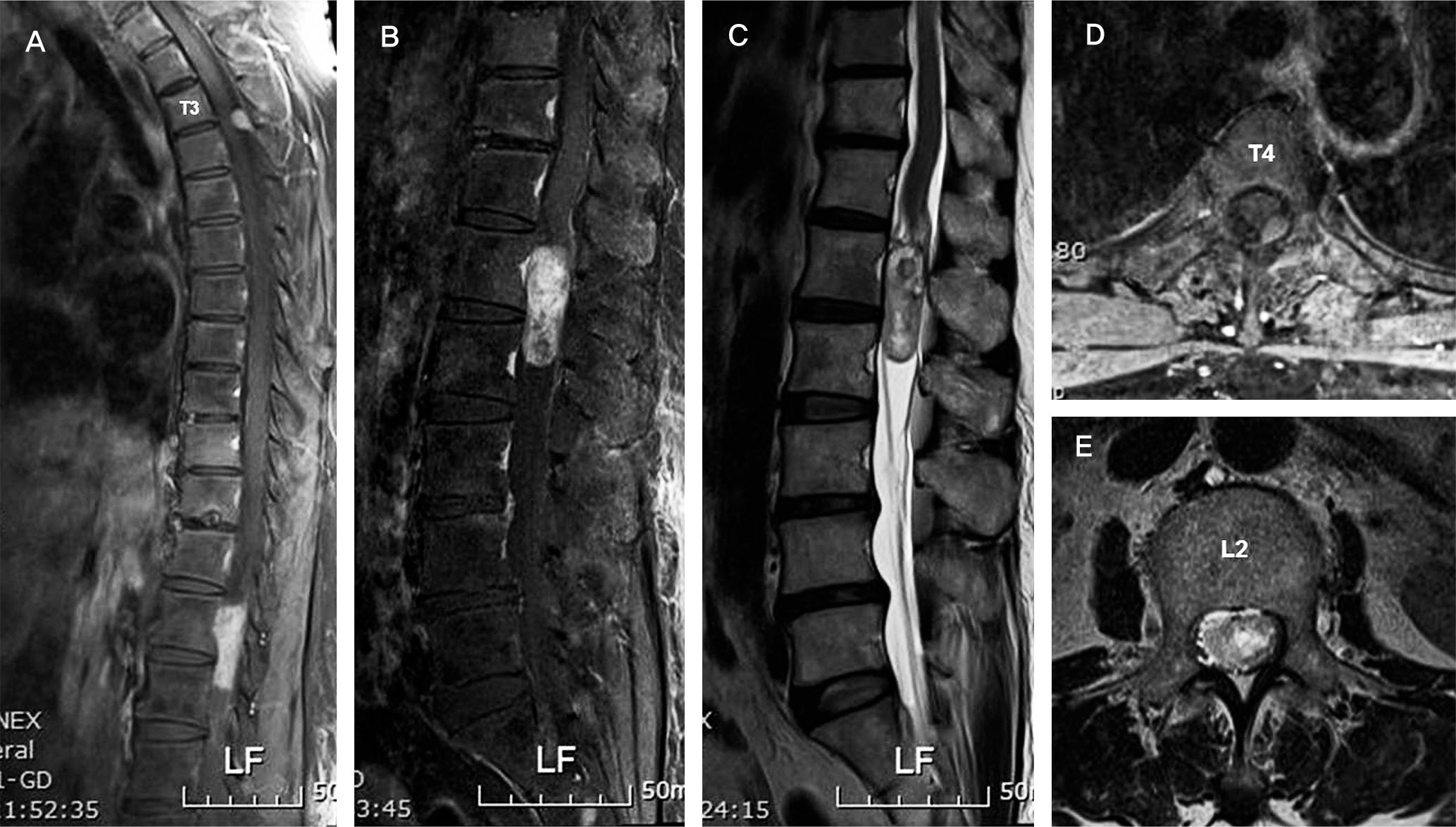
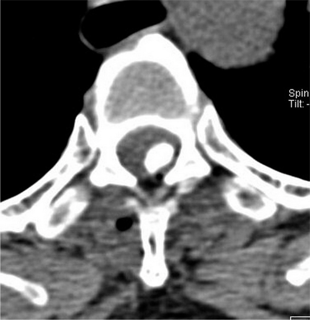
To report a case of thoracic spinal meningioma and lumbar spinal schwannoma found in one patient. SUMMARY OF LITERATURE REVIEW: patients with different types of spinal cord tumor, specifically meningioma and schwannoma, are rare in medical literature.
MATERIALS AND METHODS
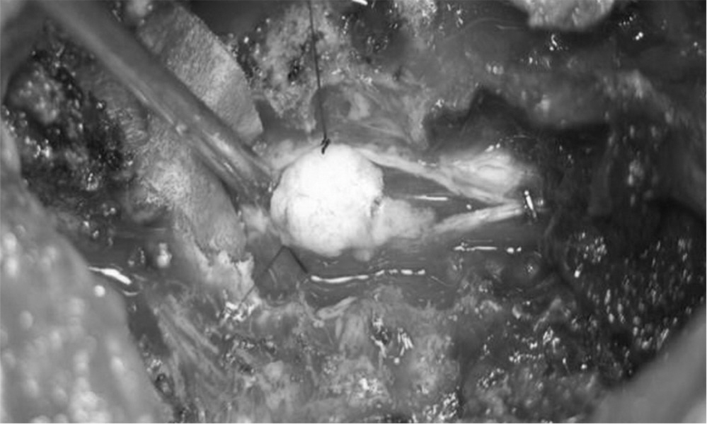
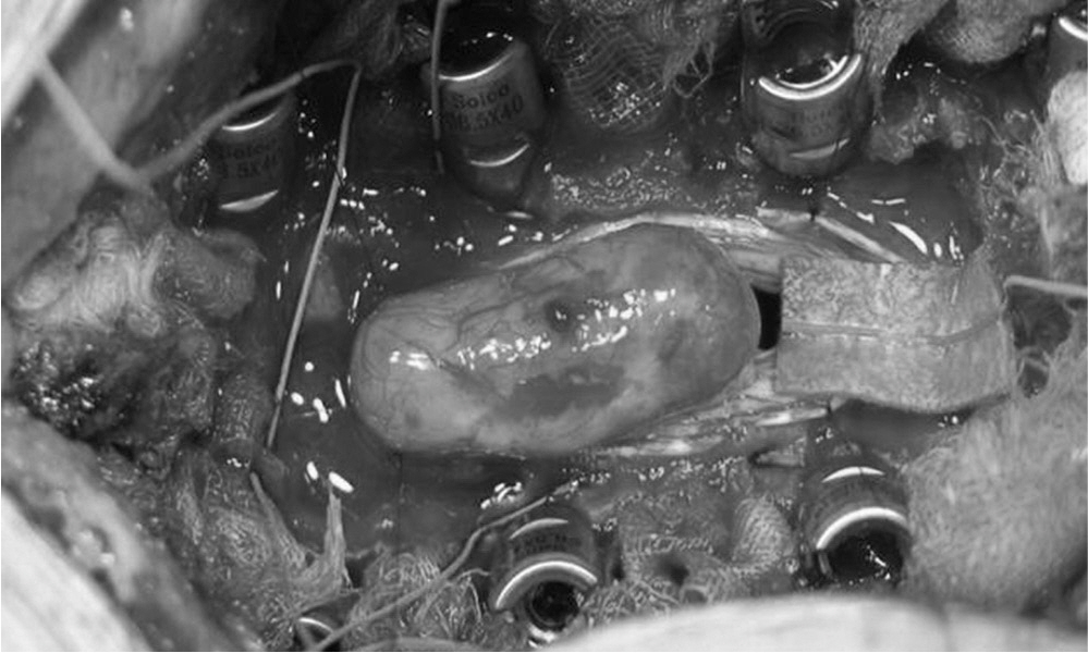
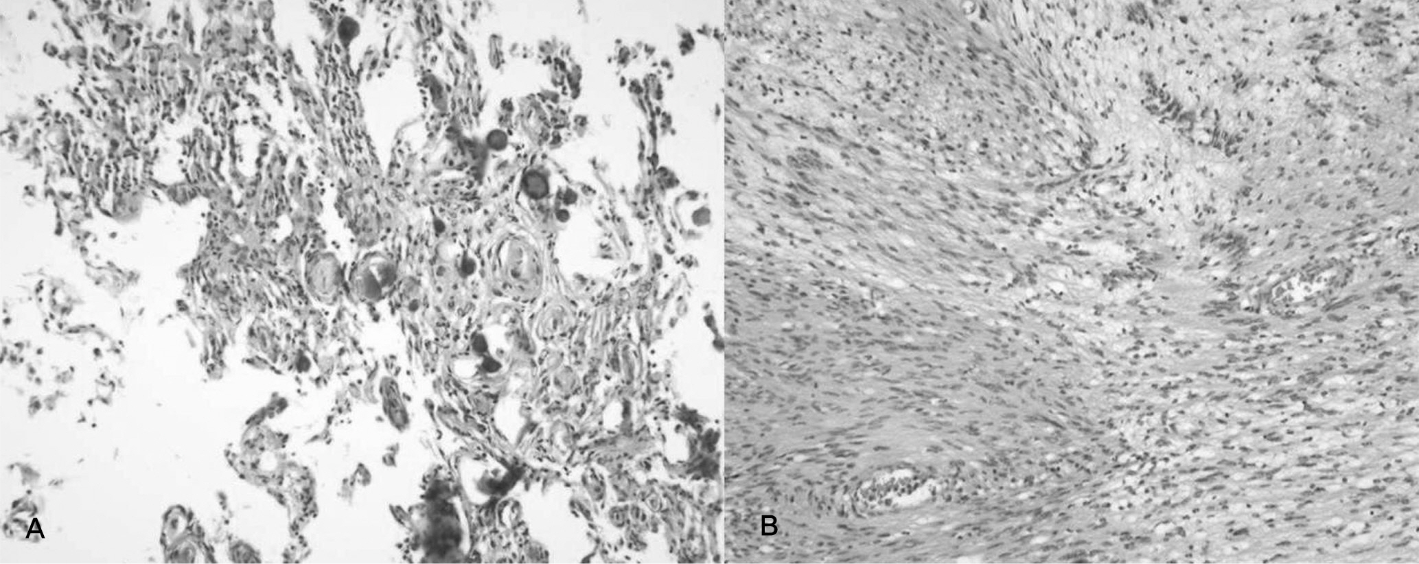
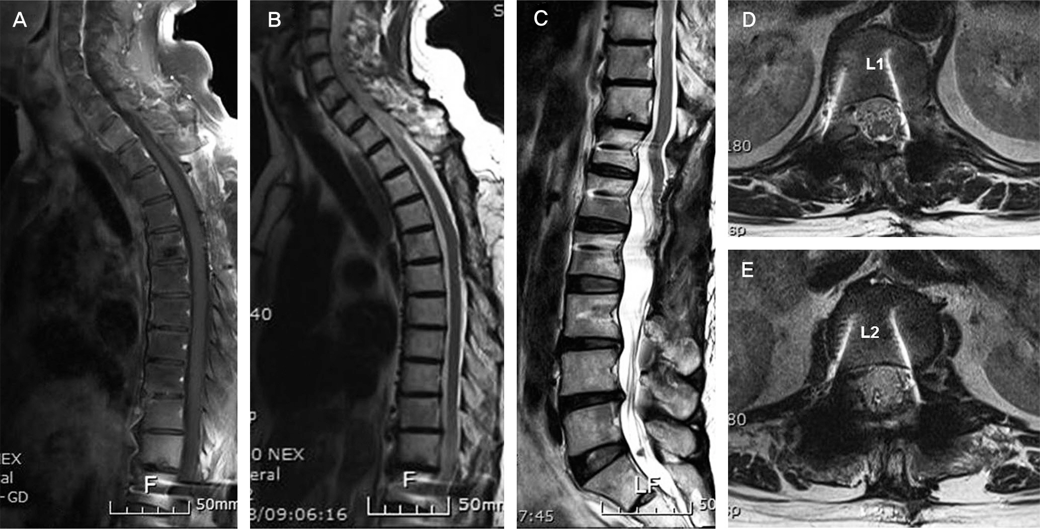
A 66 year-old female presented with complaints of walking difficulty. She had masses on the thoracic and lumbar spine and underwent open excision and biopsy.
RESULTS
Three months after operation, the patient could walk independently and no recurrence was found at 1-year follow up.
CONCLUSIONS
Thoracic spinal meningioma and lumbar spinal schwannoma occurring in one individual were treated successfully by operative management.
Keyword
Figure
Reference
-
1. Solero CL, Fornari M, Giombini S, et al. Spinal meningiomas: review of 174 operated cases. Neurosurgery. 1989; 25:153–160.
Article2. Chen HJ, Lui CC, Chen L. Spinal epidural meningioma in a child. Child's Nerv Syst. 1992; 8:465–7.
Article3. Calogero JA, Moossy J. Extradural spinal meningiomas. J Neurosurg. 1972; 37:442–7.
Article4. Nittner K. Spinal meningiomas, neurinomas, and neurofibromas and hourglass tumors. Vinken P, Brunyn B, editors. Handbook of Clinical neurology. New York: American Elsevier;1976. p. 177–322.5. Chung JY, Kim HJ, Seo HY, Lee JJ. Diverse characteristics of spinal nerve sheath tumor on magnetic resonance images. J Korean Soc Spine Surg. 2009; 16:38–45.
Article6. Bloomer CW, Ackerman A, Bhatia RG. Imaging for spinal tumors and new applications. Top Magn Reson Imaging. 2006; 17:69–87.7. Friedman DP, Tartaglino LM, Flanders AE. Intradural schwnnomas of the spine: MR findings with emphasis on contrast-enhancement characteristics. AJR. 1992; 158:1347–50.8. Demachi H, Takashima T Kadoya M, et al. MR imaging of spinal neurinomas with pathologic correlation. J Comput Assist Tomogr. 1990; 14:250–4.9. Nishiura I, Koyama T, Tanaka K, Aii H, Amano S. The occurrence of different types of spinal tumors in one patient. A case report and review of the literature. Neurochirurgia. 1989; 32:52–5.
- Full Text Links
- Actions
-
Cited
- CITED
-
- Close
- Share
- Similar articles
-
- Co-Existence of Dual Spinal Cord Tumor: Schwannoma and Meningioma
- Melanotic Schwannoma in Cervical Spine: A Case Report
- Synchronous Development of Schwannoma in the Rectus Abdominis and Lipoma in the Chest: A Case Report
- Thoracic Intramedullary Schwannoma: 2 Cases Report
- Concurrence of Schwannoma and Meningioma Neighboring in the Lumbar Spine: A Case Report

