Anat Cell Biol.
2011 Dec;44(4):324-330. 10.5115/acb.2011.44.4.324.
A simple technique for morphological measurement of cerebral arterial circle variations using public domain software (Osiris)
- Affiliations
-
- 1Department of Neurosurgery, University of Florida, Gainesville, FL, USA. saeed.ansari@neurosurgery.ufl.edu
- 2Department of Neurosurgery, Tehran University of Medical Sciences, Tehran, Iran.
- 3Department of Neurosurgery, Dunedin Hospital, Dunedin, New Zealand.
- 4Department of Anatomical Sciences, Shiraz University of Medical Sciences, Shiraz, Iran.
- KMID: 1447446
- DOI: http://doi.org/10.5115/acb.2011.44.4.324
Abstract
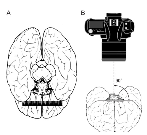
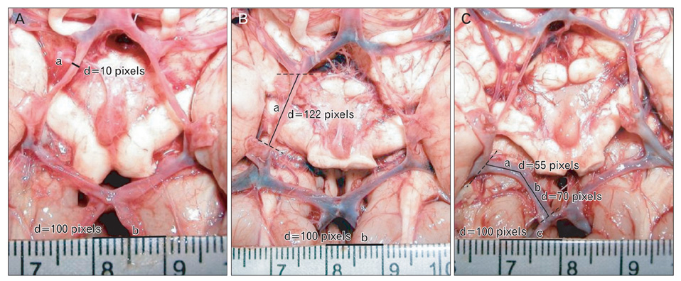
- This article describes a straightforward method to measure the dimensions and identify morphological variations in the cerebral arterial circle using the general-purpose software program Osiris. This user-friendly and portable program displays, manipulates, and analyzes medical digital images, and it has the capability to determine morphometric properties of selected blood vessels (or other anatomical structures) in humans and animals. To ascertain morphometric variations in the cerebral arterial circle, 132 brains of recently deceased fetuses, infants, and adults were dissected. The dissection procedure was first digitized, and then the dimensions were measured with Osiris software. Measurements of each vessel's length and external diameters were used to identify and classify morphological variations in the cerebral arterial circle. The most commonly observed anatomical variations were uni- and bilateral hypoplasia of the posterior communicating artery. This study demonstrates that public domain software can be used to measure and classify cerebral arterial circle vessels. This method could be extended to examine other anatomical regions or to study other animals. Additionally, knowledge of variations within the circle could be applied clinically to enhance diagnostic and treatment specificity.
MeSH Terms
Figure
Reference
-
1. Ardakani SK, Dadmehr M, Nejat F, Ansari S, Eftekhar B, Tajik P, El Khashab M, Yazdani S, Ghodsi M, Mahjoub F, Monajemzadeh M, Nazparvar B, Abdi-Rad A. The cerebral arterial circle (circulus arteriosus cerebri): an anatomical study in fetus and infant samples. Pediatr Neurosurg. 2008. 44:388–392.2. Eftekhar B, Dadmehr M, Ansari S, Ghodsi M, Nazparvar B, Ketabchi E. Are the distributions of variations of circle of Willis different in different populations? Results of an anatomical study and review of literature. BMC Neurol. 2006. 6:22.3. Lazorthes G, Gouaze A, Santini JJ, Salamon G. Le cercle artériel du cerveau (circulus arteriosus cerebri). Anat Clin. 1979. 1:241–257.4. Seydel HG. The diameters of the cerebral arteries of the human fetus. Anat Rec. 1964. 150:79–88.5. Macchi C, Catini C, Federico C, Gulisano M, Pacini P, Cecchi F, Corcos L, Brizzi E. Magnetic resonance angiographic evaluation of circulus arteriosus cerebri (circle of Willis): a morphologic study in 100 human healthy subjects. Ital J Anat Embryol. 1996. 101:115–123.6. Bhagwati SN, Deshpande HG. Study of circle of Willis in 1021 consecutive autopsies: incidence of aneurysms, anatomical variations and atherosclerosis. Ann Acad Med Singapore. 1993. 22:3 Suppl. 443–446.7. Aydın ME, Kaya AH, Kopuz C, Demir MT, Corumlu U, Dagcinar A. Bilateral origin of superior cerebellar arteries from the posterior cerebral arteries, and clues to its embryologic basis. Anat Cell Biol. 2011. 44:164–167.8. Mitchell DG, Merton DA, Mirsky PJ, Needleman L. Circle of Willis in newborns: color Doppler imaging of 53 healthy full-term infants. Radiology. 1989. 172:201–205.9. Macchi C, Giannelli F, Cecchi F, Gulisano M, Pacini P, Corcos L, Catini C, Brizzi E. The inner diameter of human intracranial vertebral artery by color Doppler method. Ital J Anat Embryol. 1996. 101:81–87.10. Macchi C, Catini C, Giannelli F, Cecchi F, Gulisano M, Pacini P, Brizzi E. The blood-flow velocity analysis as a possible method for arterial calibre discovery. A statistical investigation in 108 human subjects by color-Doppler method. Ital J Anat Embryol. 1996. 101:45–55.11. Stojanović N, Stefanović I, Randjelović S, Mitić R, Bosnjaković P, Stojanov D. Presence of anatomical variations of the circle of Willis in patients undergoing surgical treatment for ruptured intracranial aneurysms. Vojnosanit Pregl. 2009. 66:711–717.12. Rhoton AL Jr. The supratentorial arteries. Neurosurgery. 2002. 51:4 Suppl. S53–S120.13. Milenković Z, Vucetić R, Puzić M. Asymmetry and anomalies of the circle of Willis in fetal brain. Microsurgical study and functional remarks. Surg Neurol. 1985. 24:563–570.14. Saeki N, Rhoton AL Jr. Microsurgical anatomy of the upper basilar artery and the posterior circle of Willis. J Neurosurg. 1977. 46:563–578.15. Kayembe KN, Sasahara M, Hazama F. Cerebral aneurysms and variations in the circle of Willis. Stroke. 1984. 15:846–850.16. Horikoshi T, Akiyama I, Yamagata Z, Sugita M, Nukui H. Magnetic resonance angiographic evidence of sex-linked variations in the circle of Willis and the occurrence of cerebral aneurysms. J Neurosurg. 2002. 96:697–703.17. Hoksbergen AW, Majoie CB, Hulsmans FJ, Legemate DA. Assessment of the collateral function of the circle of Willis: three-dimensional time-of-flight MR angiography compared with transcranial color-coded duplex sonography. AJNR Am J Neuroradiol. 2003. 24:456–462.18. Chuang YM, Liu CY, Pan PJ, Lin CP. Posterior communicating artery hypoplasia as a risk factor for acute ischemic stroke in the absence of carotid artery occlusion. J Clin Neurosci. 2008. 15:1376–1381.19. Bugnicourt JM, Garcia PY, Peltier J, Bonnaire B, Picard C, Godefroy O. Incomplete posterior circle of willis: a risk factor for migraine? Headache. 2009. 49:879–886.20. Alpers BJ, Berry RG, Paddison RM. Anatomical studies of the circle of Willis in normal brain. AMA Arch Neurol Psychiatry. 1959. 81:409–418.21. Stock KW, Wetzel S, Kirsch E, Bongartz G, Steinbrich W, Radue EW. Anatomic evaluation of the circle of Willis: MR angiography versus intraarterial digital subtraction angiography. AJNR Am J Neuroradiol. 1996. 17:1495–1499.22. Malamateniou C, Adams ME, Srinivasan L, Allsop JM, Counsell SJ, Cowan FM, Hajnal JV, Rutherford MA. The anatomic variations of the circle of Willis in preterm-at-term and termborn infants: an MR angiography study at 3T. AJNR Am J Neuroradiol. 2009. 30:1955–1962.23. Papantchev V, Hristov S, Todorova D, Naydenov E, Paloff A, Nikolov D, Tschirkov A, Ovtscharoff W. Some variations of the circle of Willis, important for cerebral protection in aortic surgery: a study in Eastern Europeans. Eur J Cardiothorac Surg. 2007. 31:982–989.24. Van Overbeeke JJ, Hillen B, Tulleken CA. A comparative study of the circle of Willis in fetal and adult life. The configuration of the posterior bifurcation of the posterior communicating artery. J Anat. 1991. 176:45–54.25. el Khamlichi A, Azouzi M, Bellakhdar F, Ouhcein A, Lahlaidi A. Anatomic configuration of the circle of Willis in the adult studied by injection technics. Apropos of 100 brains. Neurochirurgie. 1985. 31:287–293.26. Baptista AG. Studies on the arteries of the brain. II. The anterior cerebral artery: some anatomic features and their clinical implications. Neurology. 1963. 13:825–835.27. Hillen B. The variability of the circulus arteriosus (Willisii): order or anarchy? Acta Anat (Basel). 1987. 129:74–80.28. Fisher CM. The circle of Willis: anatomical variations. Vasc Dis. 1965. 2:99–105.29. Riggs HE, Rupp C. Variation in form of circle of Willis. The relation of the variations to collateral circulation: anatomic analysis. Arch Neurol. 1963. 8:8–14.30. Orlandini GE, Ruggiero C, Orlandini SZ, Gulisano M. Blood vessel size of circulus arteriosus cerebri (circle of Willis): a statistical research on 100 human subjects. Acta Anat (Basel). 1985. 123:72–76.31. Caruso G, Vincentelli F, Rabehanta P, Giudicelli G, Grisoli F. Anomalies of the P1 segment of the posterior cerebral artery: early bifurcation or duplication, fenestration, common trunk with the superior cerebellar artery. Acta Neurochir (Wien). 1991. 109:66–71.32. Gielecki J, Zurada A, Kozłowska H, Nowak D, Loukas M. Morphometric and volumetric analysis of the middle cerebral artery in human fetuses. Acta Neurobiol Exp (Wars). 2009. 69:129–137.33. Gielecki JS, Syc B, Wilk R, Musiał-Kopiejka M, Piwowarczyk-Nowak A. Quantitative evaluation of aortic arch development using digital-image analysis. Ann Anat. 2006. 188:19–23.34. Gościcka D, Gielecki J, Materka A. Application of digital image analysis system for measurements of the blood vessels parameters (diameter, length, volume). Folia Morphol (Warsz). 1990. 49:1–7.35. Ligier Y, Ratib O, Logean M, Girard C. Osiris: a medical image-manipulation system. MD Comput. 1994. 11:212–218.36. Bird P, Lassere M, Shnier R, Edmonds J. Computerized measurement of magnetic resonance imaging erosion volumes in patients with rheumatoid arthritis: a comparison with existing magnetic resonance imaging scoring systems and standard clinical outcome measures. Arthritis Rheum. 2003. 48:614–624.37. Dulguerov P, Wang D, Perneger TV, Marchal F, Lehmann W. Videomimicography: the standards of normal revised. Arch Otolaryngol Head Neck Surg. 2003. 129:960–965.38. Hirasawa Y, Bashir WA, Smith FW, Magnusson ML, Pope MH, Takahashi K. Postural changes of the dural sac in the lumbar spines of asymptomatic individuals using positional stand-up magnetic resonance imaging. Spine (Phila Pa 1976). 2007. 32:E136–E140.
- Full Text Links
- Actions
-
Cited
- CITED
-
- Close
- Share
- Similar articles
-
- Anatomical study of variations in the configurations of the circle of Willis in relation to age, sex, and diameters of the components
- Microsurgical Study on the Circle of Willis in Korean Adults
- A Functional Perspective on the Embryology and Anatomy of the Cerebral Blood Supply
- Reduction Umbilicoplasty: A Simple and Effective Method
- Variations and anomalies of the circle of Willis in Korean: Cerebral digital subtraction angiogram studies in 200 cases


