Lab Anim Res.
2011 Dec;27(4):361-363. 10.5625/lar.2011.27.4.361.
Canine hemangiosarcoma in a female Jindo dog
- Affiliations
-
- 1College of Veterinary Medicine, Kyungpook National University, Daegu, Korea. jeongks@knu.ac.kr
- 2Stem Cell Therapeutic Research Institute, Kyungpook National University, Daegu, Korea.
- KMID: 1444972
- DOI: http://doi.org/10.5625/lar.2011.27.4.361
Abstract
- A fourteen-year female Jindo dog showing signs of hemoperitoneum was diagnosed with hemangiosarcoma through abdominal radiography and histopathological examination. In the abdominal radiographs, a circular mass was observed on the posterior of the spleen. When the splenic mass was removed via laparotomy, it was spherical, poorly circumscribed, and showed signs of necrosis, with white spots present on the outer side of the mass. Microscopically, the mass showed that hemorrhage was widespread and extensive infiltration of neoplastic cells was found throughout. There was hypercellularity, including occasional pleomorphic cells and mitotic figures. Inflammation at the edge of the mass, along with necrosis, was also found. In this report, we describe the gross and histopathological findings of a case of canine hemangiosarcoma.
Keyword
MeSH Terms
Figure
-
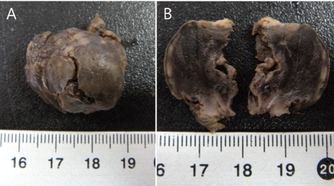
Figure 1 Gross findings in the spherical mass. (A) The spherical mass was removed from the spleen. (B) The cut surface shows white spots on the edge, indicating necrosis.
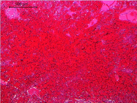
Figure 2 Hemorrhage was widespread, making it hard to recognize the normal splenic structure. H&E stain. Scale bar=500 µm.

Figure 3 Inflammation and edema were noted with the infiltration of neutrophils and the lack of cell population on the mass edge. H&E stain. Scale bar=50 µm.
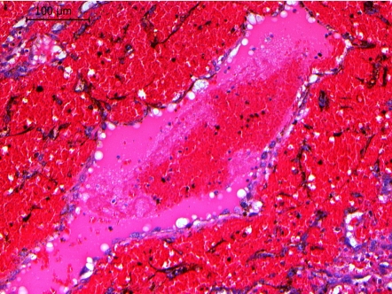
Figure 4 Multiple cavernous blood channels were formed by tumor cells. Infiltration of tumor cells and tissue reorganization were observed. H&E stain. Scale bar=100 µm.
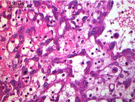
Figure 5 Scattered neoplastic cells and some mitotic figures were evident. H&E stain. Scale bar=50 µm.
Reference
-
1. Clifford CA. Treatment of canine hemangiosarcoma: 2000 and beyond. J Vet Intern Med. 2000; 14(5):479–485. PMID: 11012108.
Article2. Meuten DJ. Tumors of Domestic Animals. 2002. 4th ed. Iowa: Iowa State Press;p. 45–117.3. Mitchell BE. The Veterinary Clinics of North America, Small Animal Practice. 2003. Philadelphia: WB Saunders;p. 533–552.4. Maxie MG. Jubb, Kennedym and Palmer's Pathology of Domestic Animals. 2007. volume 3:5th ed. St. Louis: Saunders Elsevier;p. 102–105.5. Withrow SJ. Wtihrow and MacEwen's Small Animal Clinical Oncology. 2007. 4th ed. St. Louis: Saunders Elsevier;p. 785–795.6. Brown NO, Patnaik AK, MacEwen EG. Canine Hemangiosarcoma: Retrospective analysis of 104 cases. J Am Vet Med Assoc. 1985; 186:56–58. PMID: 4038395.7. Prymak C, McKee LJ, Goldschmidt MH, Glickman LT. Epidemiologic, clinical, pathologic, and prognostic characteristics of splenic hemangiosarcoma and splenic hematoma in dogs: 217 cases (1985). J Am Vet Med Assoc. 1988; 193:706–712. PMID: 3192450.
- Full Text Links
-
- Actions
-
Cited
- CITED
-
- Close
- Share
-
- Similar articles
-
- Canine behavioral problems and their effect on relinquishment of the Jindo dog
- Goniodysgenesis-associated glaucoma in a Jindo dog
- Isolation and Antimicrobial Susceptibility of Microorganisms from Milk Samples of Jindo Dogs (Canis familiaris var. jindo)
- Multiple intestinal lymphomatous polyposis in a Jindo dog
- Sebaceous adenitis in a Jindo dog: Dermatologic and histopathologic findings
